组成图示
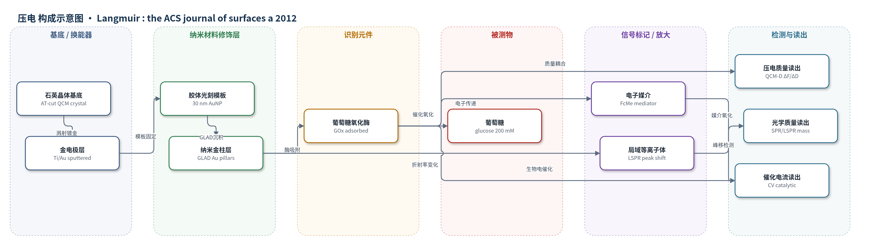
点击图片查看大图 · 依据论文自动绘制
传感器类型
压电(QCM)生物传感器
检测对象
葡萄糖氧化酶(glucose oxidase, GOx);葡萄糖(glucose);样品基质:磷酸盐缓冲液(5或20 mM,pH 7.0)
检测原理
GOx以物理吸附方式结合到平金或GLAD纳米金柱表面,识别事件使界面质量增加。在QCM-D中,吸附蛋白及其耦合水引起石英晶体频率下降和耗散变化;纳米结构内水被蛋白置换,因此需根据溶液耦合效率修正Sauerbrey质量。SPR通过平表面折射率变化独立测定干蛋白质量,LSPR则利用纳米金柱局域表面等离子体共振峰随周围折射率变化的特性,经烷硫醇校准将峰移换算为表面质量密度。电化学读出中,GOx催化葡萄糖氧化,FAD/FADH2将电子传递给FcMe,FcMe在金电极上氧化还原再生,产生与活性酶量相关的催化电流。信号随GOx吸附量和催化活性变化,可同步表征结合量、水合状态与酶功能。
检测灵敏度
—
效应效果
该方法以QCM-D、SPR、LSPR和CV交叉验证,实现纳米结构界面蛋白吸附无标记定量。纳米金表面酶负载比平表面高4–5倍,超过2.91±0.09倍表面积增幅;24 h内纳米结构表面几乎无脱附,平表面1 h后质量下降约21%。20 mM磷酸盐中吸附量高于5 mM,说明静电屏蔽影响结合。催化电流平表面为8.54±1.64 μA/cm2,纳米结构为11.3±3.4 μA/cm2;表观kcat由219±60 s−1降至90.0±29.1 s−1,提示初始部分失活。24 h后电流分别降至44.6±4.2%和55.4±6.7%,长期稳定性无显著差异。作者认为该策略适用于生物传感器酶界面表征。
传感器的构成
- 基底/换能器:AT-cut石英晶体(QCM-D sensor crystal, QSX 301)或硅片,提供压电振荡与电极支撑
- 金属电极层:溅射5 nm Ti和30 nm Au,形成导电金表面并作为SPR/QCM基底
- 模板层:PDDAC(聚二烯丙基二甲基氯化铵)处理及30 nm AuNP胶体光刻模板,控制纳米结构形貌
- 纳米修饰层:GLAD沉积Au形成纳米柱(直径49.3±6.5 nm,高65.0±5.9 nm),提供高曲率界面
- 识别元件:葡萄糖氧化酶(GOx, Aspergillus niger)直接物理吸附于金表面,催化葡萄糖氧化
- 电子媒介:FcMe(ferrocene methanol, (hydroxymethyl)ferrocene)介导GOx FAD/FADH2与电极电子转移
- 被测物:葡萄糖(glucose,200 mM)作为催化底物,用于电化学活性评估
中文摘要
纳米结构材料会显著改变吸附蛋白的行为,但相关相互作用难以表征。本文提出一种新方法,将纳米结构石英晶体微天平(QCM-D)表面的蛋白吸附研究,与表面等离子共振(SPR)和循环伏安(CV)等光学及电化学方法相结合,从而同时量化结合蛋白的量和活性。研究以氧化还原酶葡萄糖氧化酶(GOx)为模型体系,考察其在平金表面和纳米结构金表面吸附后功能行为的改变,因为该酶与材料的相互作用对生物传感器应用具有重要意义。作者采用胶体光刻和掠角沉积(GLAD)制备了曲率可控的新型纳米结构金电极表面。结果表明,即使按增大的表面积归一化,酶在纳米结构界面上的吸附量仍显著高于平界面,且24小时内未观察到明显脱附。与此同时,酶活性随时间下降,说明材料界面诱导了吸附酶的缓慢构象变化。此外,利用纳米结构材料固有的局域表面等离子体共振(LSPR)可直接量化蛋白结合。该工作展示了基于QCM-D的复杂纳米结构材料蛋白结合定量方法,可实现纳米结构界面蛋白结合的无标记检测。
英文摘要
Nanostructured materials strongly modulate the behavior of adsorbed proteins; however, the characterization of such interactions is challenging. Here we present a novel method combining protein adsorption studies at nanostructured quartz crystal microbalance sensor surfaces (QCM-D) with optical (surface plasmon resonance SPR) and electrochemical methods (cyclic voltammetry CV) allowing quantification of both bound protein amount and activity. The redox enzyme glucose oxidase is studied as a model system to explore alterations in protein functional behavior caused by adsorption onto flat and nanostructured surfaces. This enzyme and such materials interactions are relevant for biosensor applications. Novel nanostructured gold electrode surfaces with controlled curvature were fabricated using colloidal lithography and glancing angle deposition (GLAD). The adsorption of enzyme to nanostructured interfaces was found to be significantly larger compared to flat interfaces even after normalization for the increased surface area, and no substantial desorption was observed within 24 h. A decreased enzymatic activity was observed over the same period of time, which indicates a slow conformational change of the adsorbed enzyme induced by the materials interface. Additionally, we make use of inherent localized surface plasmon resonances in these nanostructured materials to directly quantify the protein binding. We hereby demonstrate a QCM-D-based methodology to quantify protein binding at complex nanostructured materials. Our approach allows label free quantification of protein binding at nanostructured interfaces.