组成图示
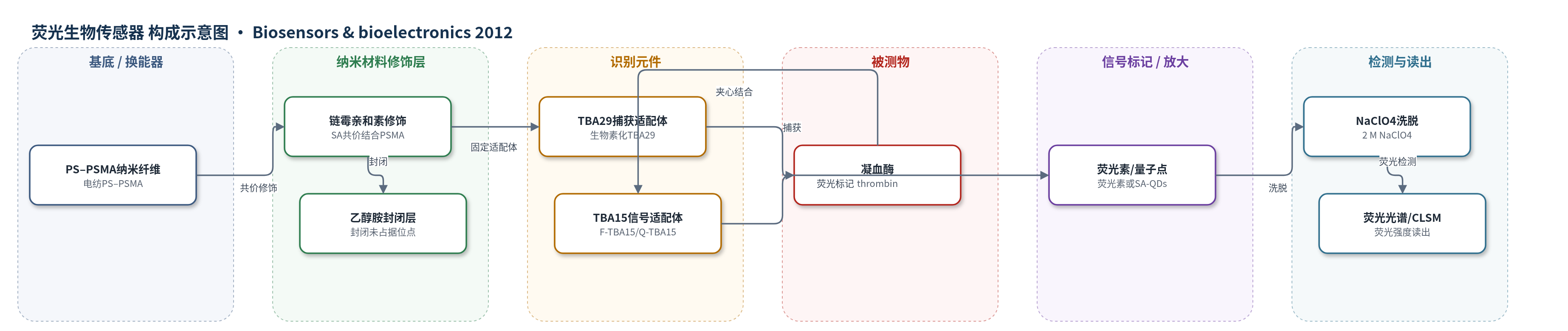
点击图片查看大图 · 依据论文自动绘制
传感器类型
荧光生物传感器
检测对象
凝血酶(thrombin);样品基质:缓冲液、10倍稀释人血清(外源加标)
检测原理
PS–PSMA纳米纤维经乙醇溶胀暴露马来酸酐基团,与链霉亲和素(SA)共价结合,再用乙醇胺封闭非特异位点;生物素化TBA29通过生物素–SA高亲和结合固定在纤维表面,作为捕获探针。加入含凝血酶的样品后,凝血酶与TBA29特异性结合并被纳米纤维捕获;洗涤去除未结合物后,加入荧光素标记TBA15(F-TBA15)或量子点标记TBA15(Q-TBA15)作为信号探针,与凝血酶另一结合位点形成夹心复合物。凝血酶浓度越高,纤维上结合的荧光标记TBA15越多。随后用2 M NaClO4将荧光标记物从纤维上洗脱至96孔板,通过荧光光谱或共聚焦显微镜测量荧光强度。量子点因高量子产率、光稳定性好,使信号放大,灵敏度优于荧光素。
检测灵敏度
LOD: 10 pM(QD标记,缓冲液);线性范围: 0.1–50 nM;R^2 = 0.99。LOD: 1 nM(荧光素标记);线性范围: 10–200 nM;R^2 = 0.99。LOD: 10 pM(QD标记,10倍稀释人血清);线性范围: 0.1–25 nM;R^2 = 0.99。
效应效果
该传感器在缓冲液中用QD标记时LOD 10 pM、线性范围0.1–50 nM(R^2=0.99),用荧光素标记时LOD 1 nM、线性范围10–200 nM(R^2=0.99);在10倍稀释人血清加标样品中仍保持LOD 10 pM,工作范围0.1–25 nM(R^2=0.99),CV 13%。选择性方面,200 nM HSA、BSA和IgG对照信号可忽略(CV 9.6%),表明SA修饰纳米纤维可减少非特异吸附。重现性方面,F-TBA15和Q-TBA15缓冲液检测平均CV分别为9.8%和11%。与96孔板相比,纳米纤维平台灵敏度提高约2500倍(96孔板Q-TBA15 LOD 25 nM、F-TBA15 LOD 50 nM)。作者认为该平台易于制备、可长期储存和重复使用,适合多种蛋白靶标检测。
传感器的构成
- 基底/载体:电纺PS–PSMA纳米纤维(PS聚苯乙烯、PSMA聚(苯乙烯-马来酸酐)),提供大比表面积三维载体
- 功能化层:链霉亲和素(SA)共价结合到PSMA马来酸酐基团,用于固定生物素化适配体
- 封闭剂:100 mM乙醇胺处理未占据区域,降低非特异结合
- 捕获识别元件:生物素化TBA29适配体(含poly dT间隔臂)通过生物素–SA结合固定,捕获凝血酶
- 被测物:凝血酶(thrombin)与TBA29特异性结合
- 信号识别元件:荧光素标记TBA15(F-TBA15)或生物素化TBA15偶联链霉亲和素量子点(SA-QDs,15–20 nm)形成Q-TBA15,结合凝血酶另一表位
- 信号读出:荧光光谱或共聚焦激光扫描显微镜(CLSM)测量洗脱后的荧光强度
中文摘要
本文报道了一种基于适配体固定的电纺聚苯乙烯–聚(苯乙烯-马来酸酐)(PS–PSMA)纳米纤维的新型适配体传感器平台,用于蛋白质检测。以两种凝血酶结合适配体TBA29和TBA15为模型,采用夹心方式检测凝血酶;适配体分别用荧光素染料或量子点标记,并通过荧光显微镜与荧光光谱测定凝血酶浓度。结果表明凝血酶可均匀捕获在纳米纤维表面。使用量子点标记时,凝血酶最低可检测浓度为10 pM,动态范围为0.1–50 nM;使用荧光素标记时,检出限为1 nM,动态范围为10–200 nM。该纳米纤维适配体传感器灵敏度比96孔板格式高约2500倍,主要归因于纳米纤维的大比表面积。此外,该平台在稀释人血清中检测外源添加凝血酶也表现出类似高灵敏度。该适配体–纳米纤维体系具有易于操作和应用的优势,有望用于多种靶标检测。
英文摘要
Here, we report the use of an aptamer-immobilized electrospun polystyrene-poly(styrene-co-maleic anhydride) (PS-PSMA) nanofiber as a new aptasensor platform for protein detection. Two thrombin-binding aptamers (TBA29 and TBA15) were used as a model platform to facilitate efficient detection of thrombin in a sandwich manner. Thrombin concentration was measured by fluorescence microscopy and spectroscopy, in which aptamers were labeled with either fluorescein dye or quantum dots. The results indicated that thrombin was captured uniformly on the surface of the nanofiber. Using this sandwich-type biosensor, the minimum detectable concentration of thrombin was 10 pM, with a dynamic range of 0.1-50 nM, when quantum dots were used for labeling. In contrast, the limit of detection was 1 nM, with a dynamic range of 10-200 nM, when using fluorescein dye labeling. This aptamers-on-nanofiber-based biosensor showed 2500-fold higher sensitivity than a 96-microwell plate format, attributed mainly to the large surface area of the nanofibers. In addition, this novel platform also exhibited similar high sensitivity in the detection of exogenously added thrombin in diluted human serum. This aptamers-on-nanofiber system, which is competitive with other sensing platforms and clinically meaningful in terms of its detection limit, is expected to be useful for the detection of various other targets because of its ease of application and manipulation.