组成图示
点击图片查看大图 · 依据论文自动绘制
传感器类型
综述或非传感器论文
检测对象
人免疫球蛋白G(human IgG, hIgG);样品基质:人血清/血清标本(文中以纯化hIgG标准品验证)
检测原理
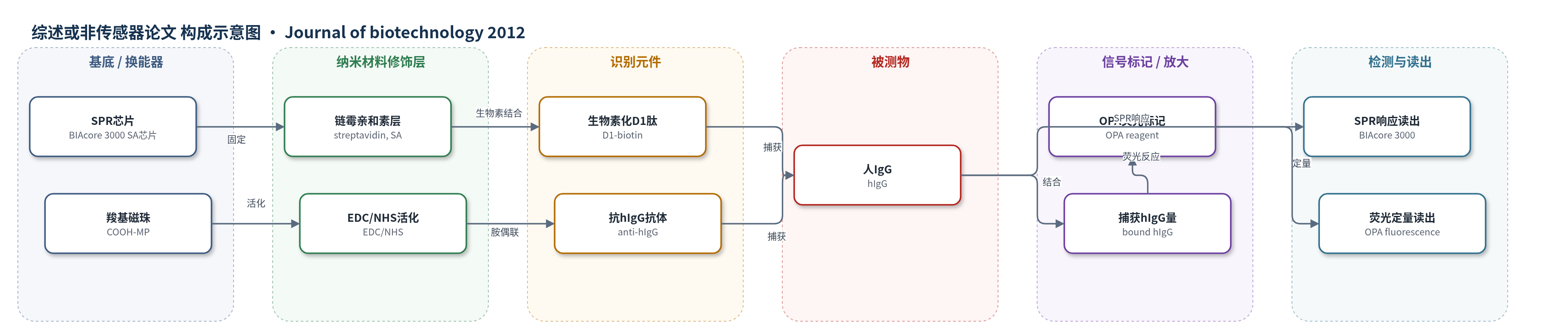
该文并非以传感器为核心,而是以亲和识别与磁分离为主。SPR表征中,生物素化抗体模拟肽或抗hIgG抗体经生物素–链霉亲和素固定于SA芯片,hIgG流过时与配体结合,引起界面折射率变化,SPR响应随hIgG浓度和结合量增大,经解离与再生可重复测定。免疫磁分离中,D1肽经生物素–链霉亲和素固定于SA-MP,或anti-hIgG抗体经EDC/NHS胺偶联固定于COOH-MP;hIgG与磁珠识别元件结合后,用磁场分离复合物。结合量用OPA荧光试剂定量,荧光强度与结合蛋白量正相关。信号依赖特异性亲和结合,未使用HCR、RCA、CRISPR-Cas或酶催化沉积等放大策略。
检测灵敏度
R^2: F9=0.9898;D1=0.9997;G5=0.9976;MA-2=0.9992;anti-hIgG=0.9796;OPA标准曲线 R^2 > 0.98
效应效果
噬菌体富集效率由1.51×10^-6升至3.98×10^-4,第2–5轮输出/输入比约增2、6、21、265倍。ELISA中12个常规和4个改良克隆结合hIgG,F9 Kd=6.21 nM,抗体0.66 nM。SPR中D1相对亲和力0.19最高。D1固定量9.3×10^-8 mol/mg-MP,抗体2.6×10^-10;捕获hIgG为1.0×10^-10和2.8×10^-10 mol/mg-MP,摩尔效率1.08×10^-3对1.08,差约1000倍。作者认为提高肽密度可补偿,肽配体成本低、稳定、简便,可用于血清hIgG去除。
传感器的构成
- 基底/换能器:BIAcore 3000 SPR芯片(SA chip),用于表面等离子共振信号读出
- 分离载体:COOH-MP/SA-MP磁珠,用于免疫磁分离
- 修饰层:链霉亲和素(streptavidin, SA)修饰SA-MP或SA chip,用于生物素化配体固定;EDC/NHS活化COOH-MP,用于抗体胺偶联
- 识别元件:生物素化12-mer抗体模拟肽D1(X12GGGS-biotin)或anti-hIgG单克隆抗体,特异性结合hIgG
- 信号标记物:OPA(o-phthalaldehyde)荧光试剂,用于结合蛋白定量;HRP-anti-M13抗体与ABTS/H2O2用于ELISA筛选
- 封闭剂:BSA/PBST用于磁珠和ELISA孔封闭,乙醇胺(ethanolamine)用于封闭COOH-MP非特异位点
- 读出层:BIAcore 3000 SPR响应、微孔板读数仪405 nm吸光度、荧光仪(330–390 nm激发/436–475 nm发射)
中文摘要
本研究以人免疫球蛋白G(hIgG)为目标抗原,通过噬菌体展示技术筛选能够模拟抗hIgG抗体结合能力的12-mer肽。实验采用常规与改良两种生物筛选(biopanning)方案,共进行5轮筛选;每轮包括将噬菌体展示12-mer肽库与hIgG包被磁珠孵育、洗涤未结合噬菌体、洗脱结合噬菌体,洗脱物可选择扩增富集或不经扩增直接进入下一轮。经ELISA筛选,鉴定出F9、D1、G5和A10等4个对hIgG具有特异性结合的克隆,其中F9的解离常数Kd为6.2 nM,仅比天然抗hIgG抗体的0.66 nM低约一个数量级。随后根据阳性克隆DNA序列化学合成4条12-mer肽,并通过SPR生物传感器测定其与hIgG的结合亲和力,结果显示D1肽具有最高亲和力。将D1肽偶联到生物功能化磁珠上,并与抗体包被磁珠比较免疫结合能力,发现肽包被磁珠的摩尔结合效率约为抗体包被磁珠的1/1000。结果表明,噬菌体展示获得的抗体模拟肽可作为亲和配体用于抗原的免疫磁分离,具有成本低、稳定性好和操作简便的潜在应用价值。
英文摘要
Phage display was performed against human IgG (hIgG) through five rounds of 'biopanning'. Each round consisted of: (1) incubating a library of phage-displayed 12-mer peptides sequences on hIgG-coated magnetic beads, (2) washing the unbound phages, and (3) eluting the bound phages. The eluted phages were either amplified to enrich the pool of positive clones or subjected to the next round without amplification. Through ELISA, four clones (F9, D1, G5, and A10) showing specific binding affinity to hIgG were identified. Among these, F9 had the highest affinity (K(d)=6.2 nM), only one order of magnitude lower than the native anti-hIgG antibody (0.66 nM). Following the DNA sequences of the selected clones, four 12-mer peptides were chemically synthesized. Among them, D1 peptide showed the highest binding affinity to hIgG via SPR biosensor measurements. This peptide was conjugated to biofunctionalized magnetic beads, and its immuno-binding ability was compared with that of the native antibody immobilized to magnetic beads. The mol-to-mol binding efficacy of the peptide-coated magnetic beads was approximately 1000-fold lower than that of the antibody-coated magnetic beads. Our results suggest a feasibility of using antibody-mimicking peptides identified by phage display technique for immuno-magnetic separation of an antigen.