组成图示
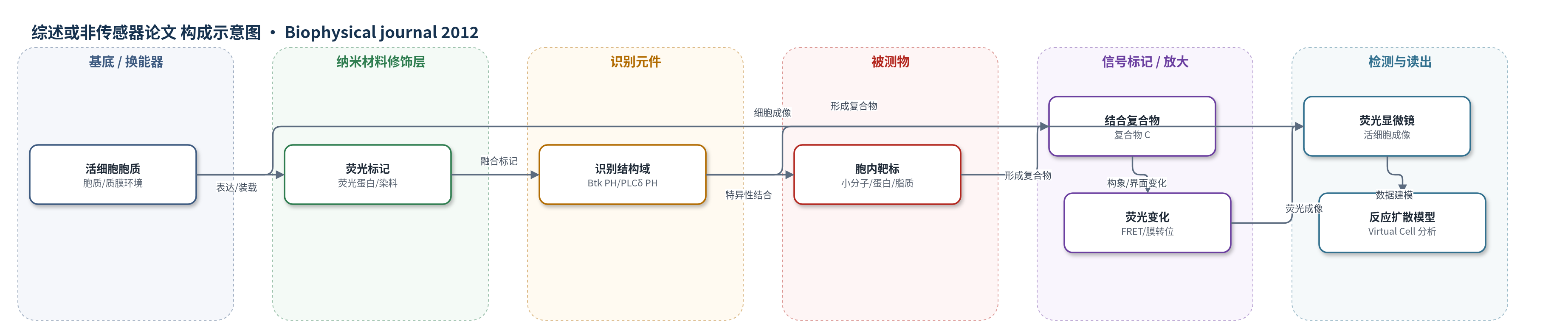
点击图片查看大图 · 依据论文自动绘制
传感器类型
综述或非传感器论文
检测对象
胞内小分子(Ca2+、cAMP)、蛋白/脂质信号分子(PtdIns(3,4,5)P3、PtdIns(4,5)P2、Ras/RhoA、蛋白激酶活性态);样品基质:活细胞胞质与质膜。
检测原理
分子生物传感器由识别元件与荧光报告基团组成。当胞内靶标(小分子、蛋白或脂质)与识别元件结合时,传感器发生构象变化、膜转位或微环境改变,使 FRET 效率、荧光强度或荧光位置发生变化;活细胞荧光显微镜将复合物 C 的局部浓度或面积密度转换为时空荧光信号。理论上信号应正比于未受扰动的自由靶标浓度,但高亲和力结合会隔离靶标,使其免受失活酶或效应因子作用,从而改变激活/失活动力学(观察者效应);若分子间传感器表达量低于靶标,未结合传感器池被耗竭,读数趋于饱和。反应-扩散模型显示,信号偏差取决于 kon/koff、靶标激活/失活速率、传感器浓度及扩散系数,空间梯度也会因结合而变缓。
检测灵敏度
原文未报告。
效应效果
本文未报告 LOD、线性范围或回收率,而是通过模型评估定量可靠性。结果表明,高亲和力识别虽能产生稳健荧光,但总会引入观察者效应或饱和效应。内分子传感器或过量分子间传感器会缓冲活性靶标,降低稳态自由靶标并减缓动力学;50% 隔离可使稳态时间约延长一倍,空间梯度变缓约 √2 倍。分子间传感器在靶标过量时读数饱和,不能反映自由靶标变化。例如 Btk PH 域结合 PtdIns(3,4,5)P3(KD=80 nM)可延长其半衰期;PLCδ PH 域结合 PtdIns(4,5)P2(KD≈2 μM)则易饱和。作者建议优化亲和力并用模型反卷积。
传感器的构成
- 样品基质/细胞环境:活细胞胞质与质膜,提供靶标存在、扩散与膜转位环境。
- 识别元件:蛋白结构域(如 Btk PH、PLCδ PH、蛋白激酶报告结构域),特异性结合胞内小分子、蛋白或脂质靶标。
- 连接元件:柔性连接肽,连接识别元件与荧光报告基团,允许结合后构象变化。
- 信号标记物:荧光蛋白或染料(FRET 供体/受体、溶剂敏感荧光基团),将结合事件转换为荧光变化。
- 读出装置:活细胞荧光显微镜,记录复合物 C 的时空分布与动力学。
中文摘要
工程化蛋白生物传感器(基于 Förster 共振能量转移、膜转位或溶剂变色位移)与活细胞荧光显微镜结合,用于实时揭示胞内过程的动力学与空间定位。该应用稳步发展,但其是否普遍适用于定量测量仍不明确。为从定量角度分析生物传感器方法的缺陷,作者分析了简单反应-扩散模型。结果表明,尽管高亲和力分子识别可稳健检测荧光读数,但会引发两种不利效应之一:内分子生物传感器或相对过量的分子间生物传感器结合会引入观察者效应,显著扰动被测靶标分子的动力学;而低表达分子间生物传感器结合则受饱和效应影响,未结合传感器池被显著耗竭。分析探讨了这些效应如何体现在生物传感器-靶标复合物的动力学和空间梯度中。令人警醒的结论是:观察者或饱和效应始终显著,问题在于能否容忍或加以校正。管理这些不利效应的挑战在于,必须将生物传感器-靶标亲和力确定到一定数量级。
英文摘要
Engineered protein biosensors, such as those based on Förster resonance energy transfer, membrane translocation, or solvatochromic shift, are being used in combination with live-cell fluorescence microscopy to reveal kinetics and spatial localization of intracellular processes as they occur. Progress in the application of this approach has been steady, yet its general suitability for quantitative measurements remains unclear. To address the pitfalls of the biosensor approach in quantitative terms, simple reaction-diffusion models were analyzed. The analysis shows that although high-affinity molecular recognition allows robust detection of the fluorescence readout, either of two detrimental effects is fostered. Binding of an intramolecular biosensor or of a relatively abundant intermolecular biosensor introduces observer effects in which the dynamics of the target molecule under study are significantly perturbed, whereas binding of a sparingly expressed intermolecular biosensor is subject to a saturation effect, where the pool of unbound biosensor is significantly depleted. The analysis explores how these effects are manifest in the kinetics and spatial gradients of the biosensor-target complex. A sobering insight emerges: the observer or saturation effect is always significant; the question is whether or not it can be tolerated or accounted for. The challenge in managing the adverse effects is that specification of the biosensor-target affinity to within a certain order of magnitude is required.