组成图示
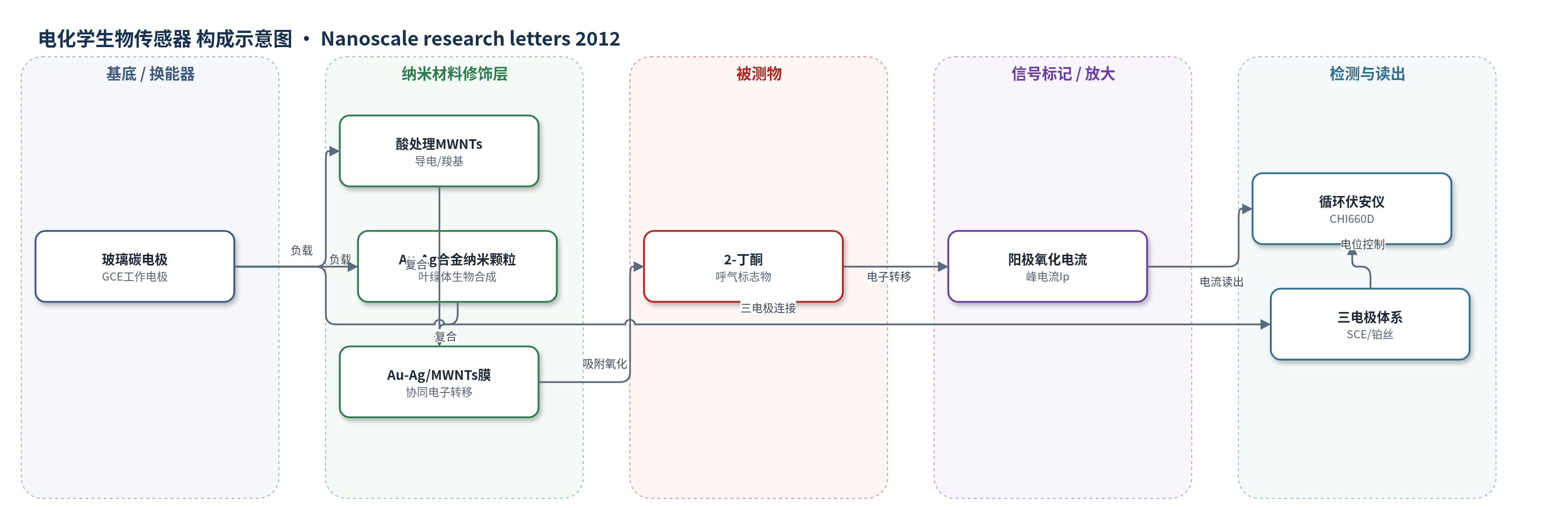
点击图片查看大图 · 依据论文自动绘制
传感器类型
电化学生物传感器
检测对象
2-丁酮(2-butanone);实验基质为 0.1 mol/L KCl 溶液,应用基质为人呼气/口腔气体
检测原理
该传感器没有引入抗体、酶或适配体等特异性识别元件,而是依靠 Au-Ag/MWNTs 纳米传感膜对 2-丁酮的直接电催化氧化。2-丁酮分子从溶液或气相样品中扩散并吸附到修饰电极表面,在循环伏安阳极扫描过程中发生氧化反应,电子经 Au-Ag 合金纳米颗粒和酸处理 MWNTs 的协同导电网络快速传递至玻璃碳电极,形成阳极电流响应。Au-Ag 合金提供催化活性位点,MWNTs 提供高导电通道,二者协同降低电子转移阻力。扫描速率实验显示峰电流与扫描速率在 20–100 mV/s 内线性相关,表明过程受吸附控制。随着 2-丁酮浓度变化,电极表面氧化反应和界面电子转移强度改变,阳极峰电流按校准曲线线性变化,从而通过循环伏安法读出浓度信息。
检测灵敏度
线性范围: 0.01%–0.075% (v/v);灵敏度斜率: -5.928×10^-6(校准方程 Ip = −5.928×10^-6 x + 1.777×10^-4);R = -0.9961
效应效果
在 0.1 mol/L KCl 中,Au-Ag/MWNTs/GCE 阳极峰电流为 220.3 μA(Ep = −0.062 V),约为 Au-Ag/GCE 的 4.2 倍,峰电位负移 31 mV,说明 MWNTs 增强电子传递。Au-Ag 与 MWNTs 比例为 1:1 时响应最高。5 个平行电极对 15 μL 2-丁酮的电流为 92.21、95.36、90.79、96.23 和 97.27 μA,RSD 3.78%(<5.0%)。KCl 溶液中保存 1 周后保留 90.4% 初始响应。论文未报告选择性、抗干扰、实际呼气样品回收率或与 GC-MS/PTR-MS 的对比,但作者认为可用于呼气或口腔气体挥发性标志物检测,具有筛查和预警早期胃癌潜力。
传感器的构成
- 基底/换能器电极:玻璃碳电极(GCE),经 0.3 和 0.05 μm 氧化铝抛光及超声清洗,作为工作电极基底。
- 纳米材料修饰层:酸处理多壁碳纳米管(MWNTs),提供导电网络,表面羧基/羰基与 Au-Ag 相互作用。
- 纳米催化层:叶绿体生物合成 Au-Ag 合金纳米颗粒(Au-Ag NPs),表面蛋白包覆,提供 2-丁酮电催化活性位点。
- 纳米传感膜:Au-Ag/MWNTs 复合膜,滴涂于 GCE 表面干燥形成传感界面,实现协同电子转移。
- 识别元件:无特异性生物识别元件,2-丁酮直接吸附并发生电催化氧化。
- 电子供体:2-丁酮(2-butanone),在电极表面氧化并向电极提供电子。
- 支持电解质:0.1 mol/L KCl 溶液,提供离子导电环境。
- 参比/辅助电极:饱和甘汞电极(SCE)和铂丝辅助电极,构成三电极电化学体系。
中文摘要
本文报道了一种一锅法、环境友好的纳米 Au-Ag 合金生物合成方法,利用叶绿体作为还原剂和稳定剂,在室温下将氯金酸和硝酸银共还原为纳米 Au-Ag 合金。通过紫外-可见吸收光谱、X 射线衍射和高分辨透射电镜对产物进行表征,傅里叶变换红外光谱进一步鉴定了叶绿体中参与合金形成与稳定的生物分子,结果表明叶绿体蛋白通过自由氨基结合到 Au-Ag 合金表面。UV-vis 中仅出现一个等离子体吸收带,说明形成的是合金结构;HR-TEM 显示颗粒呈球形,直径约 15–20 nm,SAED 和 XRD 证实其高结晶性。将所得 Au-Ag 合金分散到多壁碳纳米管中形成纳米传感膜,该膜在室温下对 2-丁酮氧化表现出强电催化活性。循环伏安法中,阳极峰电流在 0.01%–0.075%(v/v)范围内与 2-丁酮浓度呈线性关系。作者认为 Au-Ag 合金与多壁碳纳米管的协同电子转移是优异催化性能的原因,该电化学生物传感器有望用于呼气传感器,筛查和预警早期癌症,尤其是胃癌。
英文摘要
We reported a one-pot, environmentally friendly method for biosynthesizing nanoscale Au-Ag alloy using chloroplasts as reducers and stabilizers. The prepared nanoscale Au-Ag alloy was characterized by UV-visible spectroscopy, X-ray diffraction (XRD) and high resolution transmission electron microscopy (HR-TEM). Fourier transform infrared spectroscopy (FTIR) analysis was further used to identify the possible biomolecules from chloroplasts that are responsible for the formation and stabilization of Au-Ag alloy. The FTIR results showed that chloroplast proteins bound to the nanoscale Au-Ag alloy through free amino groups. The bimetallic Au-Ag nanoparticles have only one plasmon band, indicating the formation of an alloy structure. HR-TEM images showed that the prepared Au-Ag alloy was spherical and 15 to 20 nm in diameter. The high crystallinity of the Au-Ag alloy was confirmed by SAED and XRD patterns. The prepared Au-Ag alloy was dispersed into multiwalled carbon nanotubes (MWNTs) to form a nanosensing film. The nanosensing film exhibited high electrocatalytic activity for 2-butanone oxidation at room temperature. The anodic peak current (Ip) has a linear relationship with the concentrations of 2-butanone over the range of 0.01% to 0.075% (v/v), when analyzed by cyclic voltammetry. The excellent electronic catalytic characteristics might be attributed to the synergistic electron transfer effects of Au-Ag alloy and MWNTs. It can reasonably be expected that this electrochemical biosensor provided a promising platform for developing a breath sensor to screen and pre-warn of early cancer, especially gastric cancer.