组成图示
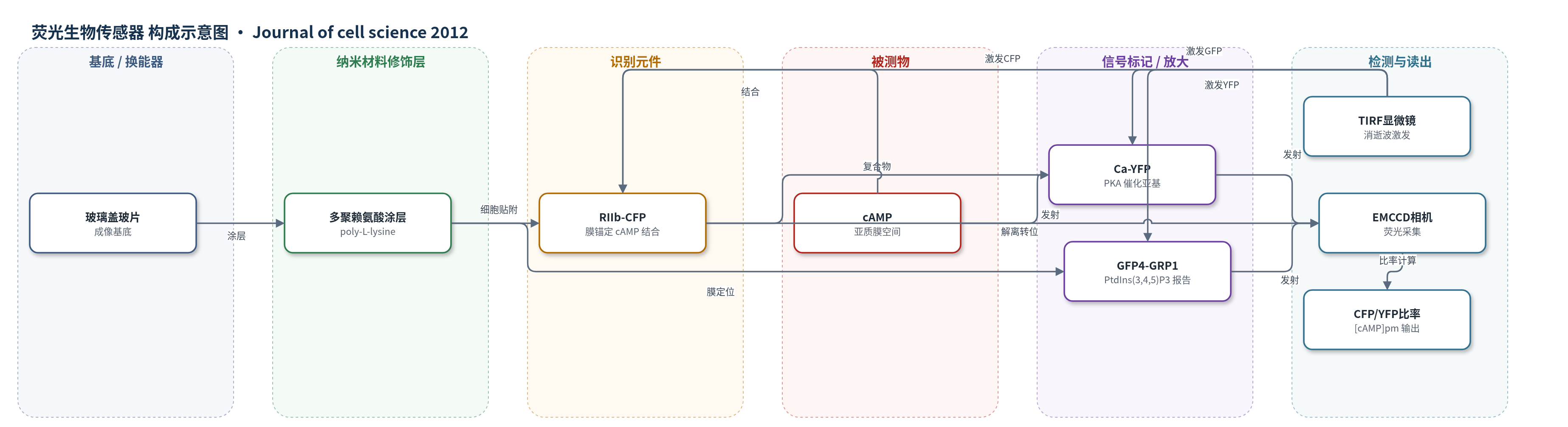
点击图片查看大图 · 依据论文自动绘制
传感器类型
荧光生物传感器
检测对象
cAMP(cyclic AMP, cAMP),样品基质为 MIN6 细胞或小鼠胰岛β细胞亚质膜空间;辅助检测 PtdIns(3,4,5)P3(磷脂酰肌醇-3,4,5-三磷酸)以反映胰岛素分泌。
检测原理
该荧光转位生物传感器由膜锚定 CFP-RIIb 与可溶性 YFP-Ca 组成。静息时 cAMP 水平低,YFP-Ca 与 RIIb-CFP 结合,CFP/YFP 比率较低。葡萄糖刺激腺苷酸环化酶生成 cAMP,cAMP 结合 RIIb 后使 Ca-YFP 从亚质膜复合物解离并扩散,CFP/YFP 比率升高。PDE1、PDE3、PDE4 和 PDE8B 水解 cAMP,决定基线、上升速率和振荡衰减;IBMX 或家族选择性抑制剂改变降解速率,从而改变 [cAMP]pm 幅度和节律。消逝波显微镜只激发膜下约百纳米区域,EMCCD 采集 CFP 与 YFP 发射,软件计算比率,实现单细胞 [cAMP]pm 时间序列读出。
检测灵敏度
原文未报告 LOD、线性范围、灵敏度斜率或相关系数。
效应效果
在 MIN6 和小鼠胰岛β细胞中,50 µM IBMX 使 55% MIN6 细胞稳定升高 [cAMP]pm,300 µM 使 81% 稳定升高;50 µM 时部分细胞出现振荡。PDE3 抑制剂 cilostamide 在静息和葡萄糖刺激下均最显著升高 [cAMP]pm,PDE1+PDE3 组合使 95% MIN6 细胞响应。葡萄糖诱导振荡幅度平均 0.27±0.02 ratio units,周期 4.7±0.4 min。PDE8B shRNA 敲低使基础 [cAMP]pm 升高,DDA 不能逆转 IBMX 升高,葡萄糖诱导振荡细胞比例从 86% 降至 45%,PtdIns(3,4,5)P3 平均荧光近加倍,胰岛素脉冲模式丢失。作者认为 PDE1、PDE3 和 PDE8B 共同塑造亚膜 cAMP 信号与脉冲胰岛素释放。
传感器的构成
- 基底/培养界面:多聚赖氨酸(poly-L-lysine)涂层玻璃盖玻片,用于 MIN6 细胞或小鼠胰岛贴附与消逝波成像。
- 光学换能层:消逝波显微镜(TIRF/evanescent wave microscopy),442 nm He-Cd 激光激发 CFP,514 nm Ar 激光激发 YFP。
- 识别元件:膜锚定截短 PKA 调节亚基 RIIb 融合 CFP(CFP-RIIb),定位亚质膜空间并结合 cAMP。
- 信号报告元件:PKA 催化亚基 Ca 融合 YFP(YFP-Ca),cAMP 结合后从 RIIb 解离并转位,改变 CFP/YFP 比率。
- 信号读出:EMCCD 相机与 MetaFluor 软件采集 CFP/YFP 比率,反映 [cAMP]pm。
- 辅助分泌报告:GFP4-GRP1 膜转位报告 PtdIns(3,4,5)P3,反映胰岛素自分泌激活。
中文摘要
环磷酸腺苷(cAMP)信号的特异性和多功能性依赖空间定位与时间动态。磷酸二酯酶(PDE)通过水解 cAMP 塑造信号。胰腺β细胞中,葡萄糖触发亚质膜 cAMP 振荡,对胰岛素分泌重要,但机制不清。本研究利用表达荧光转位生物传感器的 MIN6 细胞或小鼠胰腺β细胞,以比率式消逝波显微镜监测亚质膜 [cAMP]pm,分析不同 PDE 的作用。通用 PDE 抑制剂 IBMX 升高 [cAMP]pm;50 µM 时常见振荡,300 µM–1 mM 引起稳定升高。腺苷酸环化酶抑制剂 2',5'-二脱氧腺苷仍显著抑制 [cAMP]pm,提示存在 IBMX 不敏感降解。IBMX 敏感 PDE 中,PDE3 对维持未刺激细胞低基础 [cAMP]pm 最重要。葡萄糖诱导振荡后,PDE1、PDE3 和 PDE4 抑制剂升高平均 cAMP 水平,通常不扰乱节律。shRNA 敲低 PDE8B 升高基础 [cAMP]pm,并阻止 IBMX 存在下 DDA 的降低效应;PDE8B 敲低细胞葡萄糖诱导 [cAMP]pm 振荡减少,胰岛素脉冲分泌丢失。结论:β细胞 [cAMP]pm 振荡源于 cAMP 生成的周期性变化,PDE1、PDE3 和 PDE8B 等塑造亚膜 cAMP 信号与脉冲胰岛素释放。
英文摘要
Specificity and versatility in cyclic AMP (cAMP) signalling are governed by the spatial localisation and temporal dynamics of the signal. Phosphodiesterases (PDEs) are important for shaping cAMP signals by hydrolyzing the nucleotide. In pancreatic β-cells, glucose triggers sub-plasma-membrane cAMP oscillations, which are important for insulin secretion, but the mechanisms underlying the oscillations are poorly understood. Here, we investigated the role of different PDEs in the generation of cAMP oscillations by monitoring the concentration of cAMP in the sub-plasma-membrane space ([cAMP](pm)) with ratiometric evanescent wave microscopy in MIN6 cells or mouse pancreatic β-cells expressing a fluorescent translocation biosensor. The general PDE inhibitor IBMX increased [cAMP](pm), and whereas oscillations were frequently observed at 50 µM IBMX, 300 µM-1 mM of the inhibitor caused a stable increase in [cAMP](pm). The [cAMP](pm) was nevertheless markedly suppressed by the adenylyl cyclase inhibitor 2',5'-dideoxyadenosine, indicating IBMX-insensitive cAMP degradation. Among IBMX-sensitive PDEs, PDE3 was most important for maintaining a low basal level of [cAMP](pm) in unstimulated cells. After glucose induction of [cAMP](pm) oscillations, inhibitors of PDE1, PDE3 and PDE4 inhibitors the average cAMP level, often without disturbing the [cAMP](pm) rhythmicity. Knockdown of the IBMX-insensitive PDE8B by shRNA in MIN6 cells increased the basal level of [cAMP](pm) and prevented the [cAMP](pm)-lowering effect of 2',5'-dideoxyadenosine after exposure to IBMX. Moreover, PDE8B-knockdown cells showed reduced glucose-induced [cAMP](pm) oscillations and loss of the normal pulsatile pattern of insulin secretion. It is concluded that [cAMP](pm) oscillations in β-cells are caused by periodic variations in cAMP generation, and that several PDEs, including PDE1, PDE3 and the IBMX-insensitive PDE8B, are required for shaping the sub-membrane cAMP signals and pulsatile insulin release.