组成图示
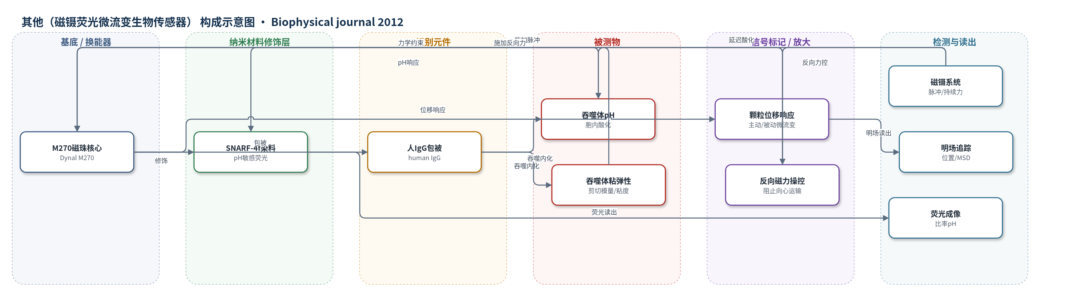
点击图片查看大图 · 依据论文自动绘制
传感器类型
其他(磁镊荧光微流变生物传感器)
检测对象
吞噬体pH(phagosomal pH)、吞噬体环境剪切模量与粘度(shear modulus and viscosity);样品基质:RAW264.7 巨噬细胞胞内吞噬体环境。
检测原理
人IgG包被的M270-SNARF颗粒被RAW264.7巨噬细胞吞噬后进入吞噬体。化学信号来自SNARF-4f:其荧光比率随吞噬体pH下降而改变,经荧光成像读出酸化动力学。力学信号来自磁性颗粒在粘弹性胞内环境中的运动:无外力时,颗粒布朗运动轨迹的均方位移按Einstein-Stokes关系换算为粘度;施加约900 pN磁脉冲时,颗粒位移响应按Voigt-Maxwell模型拟合,得到剪切模量与粘度。磁镊还可施加持续反向力,使颗粒/吞噬体位移受控,从而将局部力学状态与pH变化同步关联。方法未使用酶或核酸放大,而是依靠单颗粒磁-荧光双模态换能。
检测灵敏度
原文未报告LOD、线性范围、灵敏度斜率或相关系数。
效应效果
活细胞单颗粒同步pH与微流变测量。被动粘度0.95±0.24 Pa·s,主动粘度1±0.09×10^2 Pa·s;酸化期剪切模量45–315 Pa、粘度50–200 Pa·s。硅油对照20±3.4 Pa·s,接近22 Pa·s。磁镊可施加约4 nN、30 kT/m,操控350 nm颗粒。控制组酸化起始7.5 min,受阻组15 min,p<0.05;速率0.5±0.09 pH U/min。循环酸化受阻组15%、对照组5%,频率3.2±0.4对7.1±1.1 min。可广泛用于细胞生物物理。
传感器的构成
- 磁性换能核心:Dynal M270 磁性颗粒(Invitrogen),提供磁响应,用于磁镊受力与微流变测量
- 化学传感层:pH敏感荧光染料 SNARF-4f(Invitrogen),修饰于颗粒表面,用于比率荧光检测吞噬体pH
- 识别/吞噬配体层:人IgG(human IgG),包被颗粒表面,介导巨噬细胞吞噬受体识别与内化
- 辅助膜定位标记:K-myr-GFP(K-Ras衍生多肽-GFP),表达于质膜,用于确认吞噬杯闭合与内化时间点
- 光学读出平台:自研明场-荧光同步显微成像系统,明场追踪颗粒位置,荧光获取pH信号
- 磁镊力源:磁镊系统/磁场梯度,施加约900 pN脉冲或持续反向力,用于主动微流变与吞噬体运输操控
中文摘要
由于活细胞的化学与力学性质均发挥关键功能作用,亟需同时表征二者的生物物理方法。本文提出一种据作者所知全新的方法,利用混合磁性化学生物传感器测量局部细胞内微力学与化学性质。作者将作为化学传感器的荧光染料连接到磁性颗粒上;该颗粒通过研究其对磁脉冲的响应,用于测量周围粘弹性环境。作为概念验证,作者将该方法应用于吞噬作用研究,该过程中细胞骨架重排与吞噬体酸化并行发生。在此过程中,作者同步测量了吞噬体环境的剪切模量、粘度以及吞噬体pH。结果表明,可利用磁力使吞噬体向心运动停滞,从而操控吞噬作用。结果提示,阻止吞噬体向心运输会延迟酸化起始。据作者所知,这是首次在不干扰底层运动蛋白或细胞骨架网络、不采用生化方法的情况下,操控细胞内吞噬体运输的报道。
英文摘要
Because both the chemical and mechanical properties of living cells play crucial functional roles, there is a strong need for biophysical methods to address these properties simultaneously. Here we present a novel (to our knowledge) approach to measure local intracellular micromechanical and chemical properties using a hybrid magnetic chemical biosensor. We coupled a fluorescent dye, which serves as a chemical sensor, to a magnetic particle that is used for measurement of the viscoelastic environment by studying the response of the particle to magnetic force pulses. As a demonstration of the potential of this approach, we applied the method to study the process of phagocytosis, wherein cytoskeletal reorganization occurs in parallel with acidification of the phagosome. During this process, we measured the shear modulus and viscosity of the phagosomal environment concurrently with the phagosomal pH. We found that it is possible to manipulate phagocytosis by stalling the centripetal movement of the phagosome using magnetic force. Our results suggest that preventing centripetal phagosomal transport delays the onset of acidification. To our knowledge, this is the first report of manipulation of intracellular phagosomal transport without interfering with the underlying motor proteins or cytoskeletal network through biochemical methods.