组成图示
点击图片查看大图 · 依据论文自动绘制
传感器类型
综述或非传感器论文
检测对象
DNA损伤剂(UV、H2O2、MMS、BaP);样品基质:海胆体腔液、Aplysia dactylomela血淋巴、Panulirus argus血淋巴
检测原理
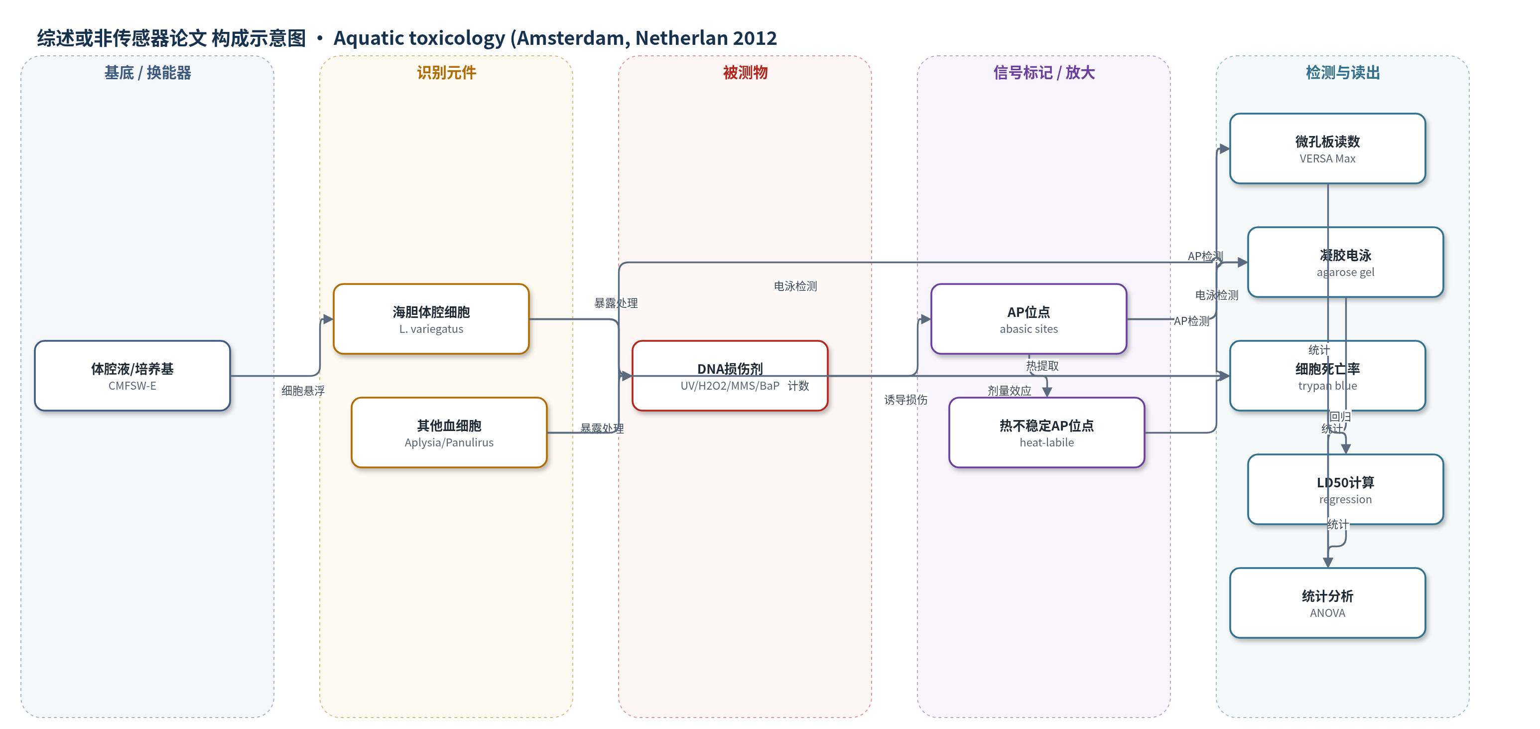
本文并非传感器检测,而是细胞基因毒性实验。将海胆体腔细胞或海洋动物血细胞暴露于不同剂量UV、H2O2、MMS和BaP,处理1、6或24 h。DNA损伤剂通过氧化脱氧核糖、烷基化碱基或形成环丁烷嘧啶二聚体等机制造成DNA损伤;部分损伤经碱基切除修复(BER)产生AP位点。随后提取DNA,用Oxiselect AP位点试剂盒在微孔板中定量AP位点,用台盼蓝排除法计数活/死细胞,并用琼脂糖凝胶电泳观察DNA断裂。信号(AP位点数、死亡率、电泳条带)随损伤剂剂量和处理时间增加而升高,但海胆体腔细胞阈值很高,仅高剂量出现显著变化。无HCR/RCA等放大策略。
检测灵敏度
未报告LOD、线性范围、灵敏度斜率、R^2。LD50(24 h):L. variegatus体腔细胞 UV 6101 ± 1019 J/m2,H2O2 120.1 ± 20.6 mM,MMS 15.4 ± 0.6 mM;A. dactylomela血细胞 UV 322 ± 20 J/m2,H2O2 2.3 ± 0.4 mM,MMS 3.3 ± 0.9 mM;P. argus血细胞 UV >9999 J/m2,H2O2 2.2 ± 0.3 mM,MMS 8.1 ± 2.0 mM。
效应效果
海胆体腔细胞对多种DNA损伤剂高度耐受。BaP最高10 μg/ml处理24 h死亡率仅8.3 ± 1.4%(n=6),未检出AP位点。UV、H2O2和MMS仅在高剂量下显著增加AP位点:UV 9999 J/m2、H2O2 300 mM 24 h、MMS 10和30 mM 6/24 h。洗涤细胞后LD50和AP位点无变化,说明抗性不依赖体腔液成分。与Aplysia dactylomela和Panulirus argus比较,Aplysia对H2O2和MMS更敏感,Panulirus对UV耐受(9999 J/m2死亡率33.2 ± 2.0%);BaP最高剂量死亡率分别为8.3 ± 1.4%、17.2 ± 2.9%和34.2 ± 6.9%。作者认为DNA损伤终点可能不适合海胆体腔细胞环境评估,但天然抗性可能与肿瘤低发相关。
传感器的构成
- 基底/换能器:未报道,本文非传感器装置,无电极或换能器。
- 纳米修饰层:未报道,无纳米材料修饰层。
- 识别元件:Lytechinus variegatus体腔细胞及Aplysia dactylomela、Panulirus argus血细胞,作为受试细胞。
- 样品基质:体腔液/血淋巴及CMFSW-E培养基,用于细胞悬浮与处理。
- 检测试剂:Oxiselect Oxidative Damage (AP sites) kit、台盼蓝、CTAB DNA提取试剂。
中文摘要
人类活动增加造成环境压力,威胁海洋生态系统。有效环境健康评估需要快速、灵敏、低成本工具预测个体和生态系统水平负面影响。为此,已开发多种基于不同细胞和生物终点的生物监测方法。海胆受精/发育试验常用于环境毒理评估,也有研究提出海胆体腔细胞可作为环境压力的新型细胞生物传感器。本研究调查了Lytechinus variegatus海胆体腔细胞对紫外线(UV)、过氧化氢(H2O2)、甲基甲磺酸(MMS)和苯并[a]芘(BaP)等DNA损伤剂的敏感性。24 h暴露后的LD50值表明体腔细胞对所有处理均高度耐受。仅在高剂量H2O2、MMS和UV下检测到DNA中脱嘌呤/脱嘧啶(AP/无碱基)位点显著增加。与Aplysia dactylomela和Panulirus argus血细胞比较显示,对不同DNA损伤剂的敏感性因物种而异。结果表明,DNA损伤可能不是海胆体腔细胞环境健康评估的有效终点,但天然抗性可能与肿瘤低发有关。
英文摘要
Increasing anthropogenic activities are creating environmental pressures that threaten marine ecosystems. Effective environmental health assessment requires the development of rapid, sensitive, and cost-effective tools to predict negative impacts at the individual and ecosystem levels. To this end, a number of biological assays using a variety of cells and organisms measuring different end points have been developed for biomonitoring programs. The sea urchin fertilization/development test has been useful for evaluating environmental toxicology and it has been proposed that sea urchin coelomocytes represent a novel cellular biosensor of environmental stress. In this study we investigated the sensitivity of coelomocytes from the sea urchin Lytechinus variegatus to a variety of DNA-damaging agents including ultraviolet (UV) radiation, hydrogen peroxide (H(2)O(2)), methylmethane sulfonate (MMS) and benzo[a]pyrene (BaP). LD(50) values determined for coelomocytes after 24h of exposure to these DNA damaging agents indicated a high level of resistance to all treatments. Significant increases in the formation of apurinic/apyrimidinic (AP or abasic) sites in DNA were only detected using high doses of H(2)O(2), MMS and UV radiation. Comparison of sea urchin coelomocytes with hemocytes from the gastropod mollusk Aplysia dactylomela and the decapod crustacean Panulirus argus indicated that sensitivity to different DNA damaging agents varies between species. The high level of resistance to genotoxic agents suggests that DNA damage may not be an informative end point for environmental health assessment using sea urchin coelomocytes however, natural resistance to DNA damaging agents may have implications for the occurrence of neoplastic disease in these animals.