组成图示
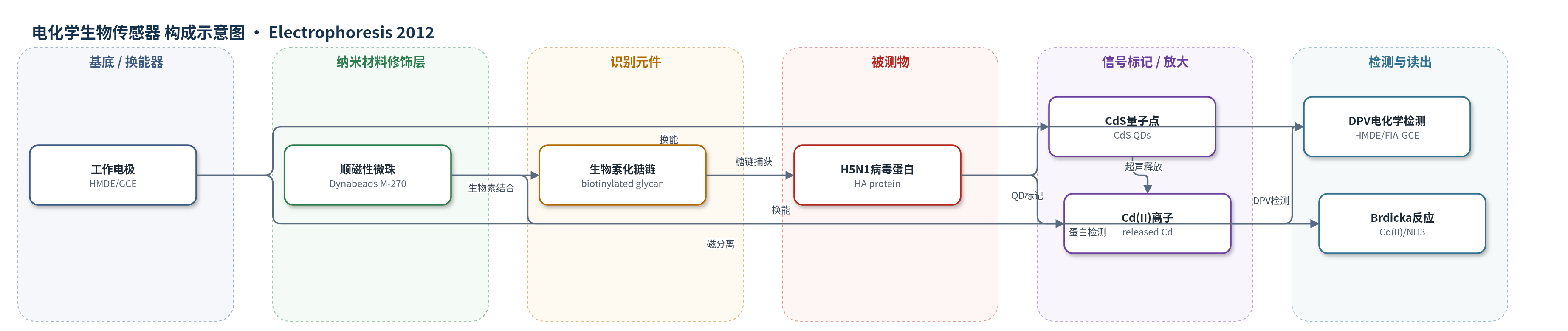
点击图片查看大图 · 依据论文自动绘制
传感器类型
电化学生物传感器
检测对象
流感病毒血凝素蛋白(Hemagglutinin, HA,A/H5N1/Vietnam/1203/2004);样品基质:病毒蛋白溶液/标准品
检测原理
该检测以生物素化糖链修饰链霉亲和素顺磁性微珠作为识别界面。生物素-链霉亲和素作用将糖链固定于磁珠表面,糖链末端唾液酸与流感病毒血凝素(HA)特异性结合,从而捕获预先用CdS量子点标记的H5N1蛋白。超声处理使捕获复合物从磁珠上释放,CdS QDs中的Cd(II)进入溶液;在HMDE或FIA-GCE上,Cd(II)经差分脉冲伏安法还原/溶出,峰高随Cd(II)量增加,间接反映蛋白浓度。另一路径中,蛋白巯基在Brdicka反应中与Co(II)在汞电极上催化产氢,Cat2峰高随蛋白浓度增加。量子点携带多个Cd(II),提供电化学信号放大。
检测灵敏度
LOD: 0.1 μg/mL (HMDE, Cd-based);LOD: 10 μg/mL (GCE, abstract);LOD: 1 μg/mL (FIA-GCE, results);LOD: 0.1 μg/mL (Brdicka reaction, abstract);LOD: 100 ng/mL (Brdicka reaction, results);线性范围: 0.1–100 μM Cd(II) (HMDE);线性范围: 0.25–30 μM Cd(II) (HMDE/GCE correlation);线性范围: 0.625–5 μg/mL H5N1-CdS (optimized assay);斜率: 1.6958 (HMDE Cd);斜率: 0.7710 (FIA-GCE Cd);斜率: 0.2871 (HMDE H5N1-CdS);斜率: 0.0284 (optimized HMDE);斜率: 0.024 (optimized FIA-GCE);R^2 = 0.9911 (HMDE Cd);R^2 = 0.9982 (FIA-GCE Cd);R^2 = 0.9973 (HMDE H5N1-CdS);R^2 = 0.9904 (optimized HMDE);R^2 = 0.9923 (optimized FIA-GCE);RSD = 4.8 (HMDE Cd);RSD = 6.3 (FIA-GCE Cd);RSD = 2.1% (HMDE H5N1-CdS);RSD = 4.8% (FIA-GCE H5N1-CdS);detectable Cd: 10 ng/mg protein
效应效果
体系在优化条件(250 μg/mL糖链、30 min、25°C、400 rpm)下选择性捕获H5N1-CdS复合物。CdS QDs稳定性超过1个月。Cd(II)检测在HMDE上RSD为4.8,FIA-GCE上RSD为6.3;H5N1-CdS复合物在HMDE上RSD为2.1%,FIA-GCE上RSD为4.8%。每个复合物约含20个Cd(II),蛋白中可检测Cd含量为10 ng/mg,支持亚微克级蛋白识别。未报告实际样品加标回收率。作者认为该自动化FIA-磁分离-电化学方法成本低、速度快,适合流感病毒蛋白的大规模筛查和分散式检测。
传感器的构成
- 换能器电极:HMDE(悬挂汞滴电极,工作电极,面积0.4 mm²)或GCE(玻璃碳电极,FIA流通池工作电极),配合Ag/AgCl/3M KCl参比电极和Pt/钢管辅助电极,用于DPV信号读出
- 磁分离载体:Streptavidin Dynabeads M-270(链霉亲和素修饰顺磁性微珠),用于固定识别元件并实现磁分离
- 识别元件:biotinylated multivalent glycans(生物素化多价糖链,Neu5Acα2–3Galβ1–3GlcNAcβ1-PAA-biotin),通过生物素-链霉亲和素结合固定于磁珠,选择性结合流感HA蛋白
- 信号标记物:CdS QDs(硫化镉量子点,3-mercaptopropionic acid修饰),与H5N1病毒蛋白形成复合物,提供Cd(II)电化学信号
- 洗脱介质:0.3 M磷酸盐缓冲液(pH 7.4)与超声针处理,用于释放捕获的蛋白/CdS QDs复合物
- 支持电解质:0.2 M醋酸缓冲液(pH 5)用于Cd(II) DPV;Brdicka支持电解质(1 mM Co(NH3)6Cl3 + 1 M氨缓冲液,pH 9.6)用于蛋白Brdicka反应
- 自动化流路:FIA系统(eVol注射泵、Valco阀、CH流通池、Metrohm 910 PSTAT mini),用于自动进样和电化学检测
中文摘要
本研究报道了一种基于流动注射分析(FIA)的电化学生物传感器,用于快速、低成本检测流感病毒蛋白。该体系以链霉亲和素修饰的顺磁性微珠为载体,表面结合生物素化多价糖链,利用糖链与流感病毒血凝素(HA)的特异性结合,选择性捕获用硫化镉量子点(CdS QDs)标记的 A/H5N1/Vietnam/1203/2004 病毒蛋白。捕获后经超声释放,采用差分脉冲伏安法(DPV)在悬挂汞滴电极(HMDE)和/或玻璃碳电极(GCE)上定量 Cd(II) 离子,从而间接测定病毒蛋白;同时用 Brdicka 反应直接检测蛋白。基于 Cd(II) 定量的检测限在 HMDE 和 GCE 上分别为 0.1 μg/mL 和 10 μg/mL;Brdicka 反应检测限为 0.1 μg/mL。优化条件下可识别亚微克级病毒蛋白。
英文摘要
Currently, the influenza virus infects millions of individuals every year. Since the influenza virus represents one of the greatest threats, it is necessary to develop a diagnostic technique that can quickly, inexpensively, and accurately detect the virus to effectively treat and control seasonal and pandemic strains. This study presents an alternative to current detection methods. The flow-injection analysis-based biosensor, which can rapidly and economically analyze a wide panel of influenza virus strains by using paramagnetic particles modified with glycan, can selectively bind to specific viral A/H5N1/Vietnam/1203/2004 protein-labeled quantum dots. Optimized detection of cadmium sulfide quantum dots (CdS QDs)-protein complexes connected to paramagnetic microbeads was performed using differential pulse voltammetry on the surface of a hanging mercury drop electrode (HMDE) and/or glassy carbon electrode (GCE). Detection limit (3 S/N) estimations based on cadmium(II) ions quantification were 0.1 μg/mL or 10 μg/mL viral protein at HMDE or GCE, respectively. Viral protein detection was directly determined using differential pulse voltammetry Brdicka reaction. The limit detection (3 S/N) of viral protein was estimated as 0.1 μg/mL. Streptavidin-modified paramagnetic particles were mixed with biotinylated selective glycan to modify their surfaces. Under optimized conditions (250 μg/mL of glycan, 30-min long interaction with viral protein, 25°C and 400 rpm), the viral protein labeled with quantum dots was selectively isolated and its cadmium(II) content was determined. Cadmium was present in detectable amounts of 10 ng per mg of protein. Using this method, submicrogram concentrations of viral proteins can be identified.