组成图示
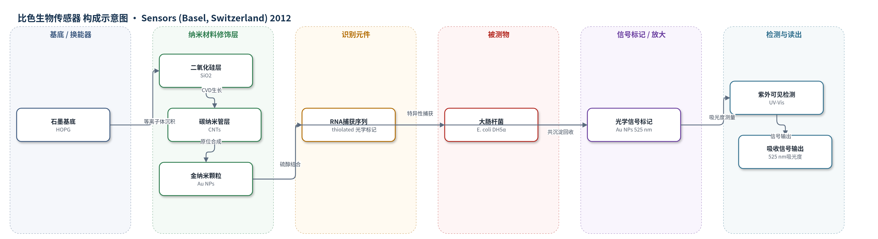
点击图片查看大图 · 依据论文自动绘制
传感器类型
比色生物传感器
检测对象
大肠杆菌(Escherichia coli,DH5α菌株);样品基质:磷酸盐缓冲液(PBS)细胞悬液,作者展望环境水样
检测原理
该传感器以碳纳米管/金纳米颗粒复合界面为捕获平台,硫醇化RNA通过Au-S键固定于金纳米颗粒表面,RNA序列特异性识别大肠杆菌表面分子。细菌结合后,金纳米颗粒随细胞沉淀回收,未结合颗粒被去除;回收样品中金纳米颗粒的局域表面等离子共振(LSPR)吸收峰位于约525 nm,吸光度与捕获的金纳米颗粒数量成正比,从而间接反映大肠杆菌浓度。碳纳米管保留电学特性并提高界面面积,金纳米颗粒提供额外结合位点,但当前未使用HCR、RCA、CRISPR-Cas或酶催化沉积等信号放大策略。
检测灵敏度
未报告 LOD、线性范围、灵敏度斜率或相关系数。
效应效果
该研究以三次重复实验验证RNA修饰效果。与裸金纳米颗粒相比,RNA包覆金纳米颗粒对大肠杆菌的捕获量提高189%,表明RNA序列具有选择性识别作用。动态光散射和Zeta电位显示,金纳米颗粒结合RNA后水合粒径由44.0 nm增至55.0 nm,表面电荷由-41.8 mV变为-32.8 mV,证实RNA固定。完整传感器经SEM观察可结合大肠杆菌,但裸传感器与RNA修饰传感器间无显著差异,作者推测RNA与碳纳米管π-π相互作用导致RNA折叠,降低可用结合位点。论文未报告稳定性、RSD、实际样品回收率或与ELISA/HPLC/qPCR的对比。作者认为该平台可拓展为多阵列电化学传感器,用于环境病原体快速检测。
传感器的构成
- 基底:高定向热解石墨(HOPG)/石墨片,表面等离子体衍生二氧化硅(SiO2)薄层,提供耐高温、化学惰性支撑并促进碳纳米管生长
- 碳纳米管层:碳纳米管(CNTs),通过化学气相沉积(CVD)在SiO2/石墨上高密度生长,提供导电网络、高比表面积和电学换能基础
- 金纳米颗粒层:金纳米颗粒(Au NPs),由HAuCl4、柠檬酸钠和NaBH4在CNT表面原位合成,粒径约44.9±14.4 nm,增加表面积并提供硫醇结合位点
- 识别元件:硫醇化RNA捕获序列(thiolated RNA capture element/aptamer),基于SELEX获得,特异性识别DH5α大肠杆菌表面,通过3'端硫醇与Au NPs结合
- 信号标记物:金纳米颗粒(Au NPs)作为光学标记,其局域表面等离子共振吸收峰约525 nm,用于UV-Vis比色读出
- 读出:紫外-可见分光光度计(UV-Vis)测量回收细胞中Au NPs在约525 nm的吸收强度,间接反映大肠杆菌捕获量
中文摘要
本研究考察了一种纳米特征生物传感器平台的构建,旨在快速、选择性检测大肠杆菌。该传感器以碳纳米管为基底,其表面修饰金纳米颗粒,并在金纳米颗粒上固定一种特异性表面黏附RNA序列元件。多步传感器组装通过以下步骤完成:在石墨基底上生长碳纳米管,在碳纳米管表面直接合成金纳米颗粒,以及将硫醇化RNA结合到已固定的金纳米颗粒上。将复合纳米材料用于传感器开发具有明显优势:既保留碳纳米管的电学行为,又通过金纳米颗粒提供更大表面积,增加分析物结合位点,从而提高灵敏度。研究成功证明,与未修饰颗粒相比,用选择性RNA序列包覆金纳米颗粒可使大肠杆菌捕获量提高189%。本文所述的传感器构建方法表明,独特复合结构在开发用于快速、灵敏检测病原体的多阵列电化学传感器方面具有巨大潜力。
英文摘要
This study examines the creation of a nano-featured biosensor platform designed for the rapid and selective detection of the bacterium Escherichia coli. The foundation of this sensor is carbon nanotubes decorated with gold nanoparticles that are modified with a specific, surface adherent ribonucleiuc acid (RNA) sequence element. The multi-step sensor assembly was accomplished by growing carbon nanotubes on a graphite substrate, the direct synthesis of gold nanoparticles on the nanotube surface, and the attachment of thiolated RNA to the bound nanoparticles. The application of the compounded nano-materials for sensor development has the distinct advantage of retaining the electrical behavior property of carbon nanotubes and, through the gold nanoparticles, incorporating an increased surface area for additional analyte attachment sites, thus increasing sensitivity. We successfully demonstrated that the coating of gold nanoparticles with a selective RNA sequence increased the capture of E. coli by 189% when compared to uncoated particles. The approach to sensor formation detailed in this study illustrates the great potential of unique composite structures in the development of a multi-array, electrochemical sensor for the fast and sensitive detection of pathogens.