组成图示
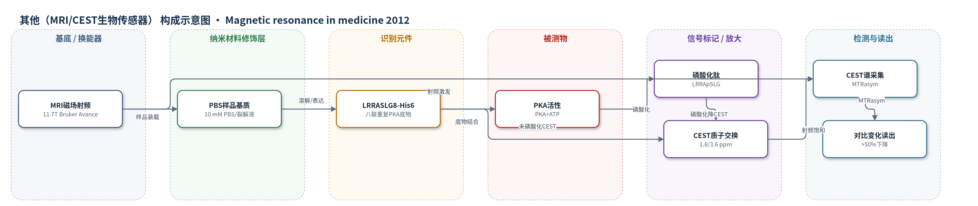
点击图片查看大图 · 依据论文自动绘制
传感器类型
其他(MRI/CEST生物传感器)
检测对象
蛋白激酶A活性(protein kinase A, PKA)/PKA介导的磷酸化;样品基质:10 mM PBS、E. coli细胞裂解液
检测原理
该传感器以PKA天然底物肽LRRASLG为识别元件,其精氨酸胍基和酰胺质子与水质子发生化学交换,在1.8和3.6 ppm处产生CEST对比。CEST-MRI用射频脉冲饱和这些质子,饱和经化学交换转移至水质子,使水质子信号下降,并以MTRasym读出。当PKA存在时,PKA利用ATP将LRRASLG中丝氨酸磷酸化;磷酸基负电荷改变邻近胍基/酰胺质子交换速率,并消除丝氨酸羟基质子,导致CEST对比下降。八联重复LRRASLG8-His6通过多个重复单元放大CEST信号。PKA活性越高,磷酸化比例越高,CEST峰下降越明显,从而实现分钟级、浓度依赖的PKA活性检测。
检测灵敏度
原文未报告LOD、线性范围、灵敏度斜率、R^2。
效应效果
体外实验中,未磷酸化LRRASLG在1.8和3.6 ppm处产生明显CEST峰,磷酸化后MTRasym对比下降超过50%,且该变化在1 mM浓度下呈浓度依赖性,可至微摩尔水平。PKA孵育时间曲线显示,大部分CEST对比变化在前15 min内产生,与重组PKA活性动力学一致。E. coli表达LRRASLG8-His6的裂解液相比表达对照蛋白胞嘧啶脱氨酶的裂解液,在1.8和3.6 ppm处MTRasym更高,说明基因编码传感器可在生物学相关环境中被检测。作者认为该MRI生物传感器可用于非侵入监测小分子或细胞治疗反应,并可拓展至PKC、CaMKIIa等蛋白激酶。
传感器的构成
- 换能器/检测场:11.7 T Bruker Avance MRI系统,提供射频饱和与磁场,用于CEST信号读出
- 样品基质:10 mM PBS(pH 7.4)或E. coli裂解液,承载传感器肽/蛋白
- 识别元件:LRRASLG肽(PKA天然底物),丝氨酸残基被PKA磷酸化
- 信号放大元件:LRRASLG8-His6蛋白(6.8 kDa,八联重复),增强CEST对比并便于纯化
- 信号转换元件:Ser-OH及邻近Arg胍基/酰胺质子,磷酸化后改变质子交换速率
- 反应底物/酶源:PKA与ATP,催化LRRASLG磷酸化
中文摘要
蛋白激酶(包括蛋白激酶A,PKA)参与众多关键细胞信号通路,但目前在遗传指定细胞群体中实时、高空间分辨率地监测激酶活性仍是未满足需求,对理解复杂生物系统及新药筛选至关重要。作者基于蛋白激酶天然识别序列可通过化学交换饱和转移磁共振成像(CEST-MRI)检测的假设,设计了一种基因编码生物传感器,其由PKA天然底物肽LRRASLG的八个串联重复组成。结果显示,该传感器在PKA磷酸化后出现可测量的CEST信号变化。天然PKA底物LRRASLG在1.8和3.6 ppm(摘要原文为11.8和13.6 ppm)处具有CEST-MRI对比,磷酸化后对比变化超过50%,并具备分钟级时间分辨率。表达编码八个LRRASLG单体的合成基因后,在相应CEST频率处出现两个峰。综上,该基因可用于在生物学相关体系中检测PKA水平,且该设计策略可拓展至多种临床相关蛋白激酶。
英文摘要
PURPOSE: Protein kinases including protein kinase A (PKA) underlie myriad important signaling pathways. The ability to monitor kinase activity in vivo and in real-time with high spatial resolution in genetically specified cellular populations is a yet unmet need, crucial for understanding complex biological systems as well as for preclinical development and screening of novel therapeutics.
METHODS: Using the hypothesis that the natural recognition sequences of protein kinases may be detected using chemical exchange saturation transfer magnetic resonance imaging, we designed a genetically encoded biosensor composed of eight tandem repeats of the peptide LRRASLG, a natural target of PKA.
RESULTS: This sensor displays a measurable change in chemical exchange saturation transfer signal following phosphorylation by PKA. The natural PKA substrate LRRASLG exhibits a chemical exchange saturation transfer-magnetic resonance imaging contrast at +1.8 and +3.6 ppm, with a >50% change after phosphorylation with minutes-scale temporal resolution. Expression of a synthetic gene encoding eight monomers of LRRASLG yielded two peaks at these chemical exchange saturation transfer frequencies.
CONCLUSION: Taken together, these results suggest that this gene may be used to assay PKA levels in a biologically relevant system. Importantly, the design strategy used for this specific sensor may be adapted for a host of clinically interesting protein kinases.