组成图示
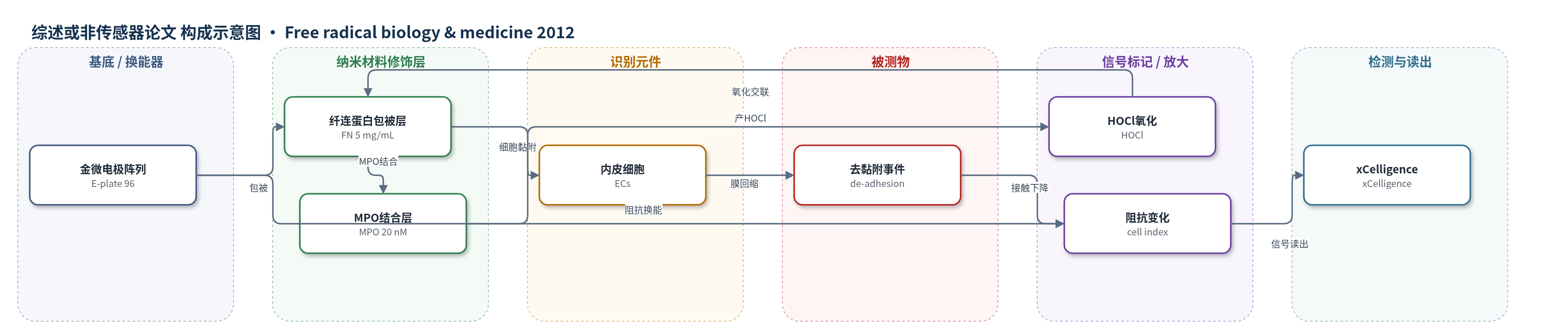
点击图片查看大图 · 依据论文自动绘制
传感器类型
综述或非传感器论文
检测对象
内皮细胞去黏附(endothelial cell de-adhesion);样品基质:培养牛主动脉内皮细胞(ECs)单层
检测原理
该检测基于实时细胞-基底阻抗。金微电极阵列上包被纤连蛋白(FN),髓过氧化物酶(MPO)结合于 FN。加入低微摩尔 H2O2 后,MPO 催化 Cl- 氧化生成高反应性 HOCl;HOCl 在 FN 附近局部氧化并共价交联 FN,破坏其细胞黏附位点。内皮细胞(ECs)通过整合素/黏附斑与 FN 结合,FN 功能受损后细胞-基质接触减弱,细胞膜从基底和邻近细胞回缩,形成去黏附。细胞-基底接触面积下降改变金电极界面的交流阻抗,xCelligence 系统将其转换为 cell index 下降;去黏附程度越大,cell index 下降越明显。MPO 催化将 H2O2 转化为 HOCl,形成局部氧化放大;预存肌动球蛋白张力进一步加速膜回缩,使信号快速出现。
检测灵敏度
原文未报告 LOD、线性范围、灵敏度斜率或相关系数。
效应效果
MPO 结合 FN 本身不改变 EC 黏附,但 MPO-FN 表面经 1–10 μM H2O2 预处理后,EC 黏附和铺展显著下降;该效应可被 Met、ABAH、SCN- 或 NO2- 减轻,说明依赖 HOCl。含 MPO 的 EC 在 5–25 μM H2O2 处理下出现快速、剂量依赖的 cell index 下降,2 min 内即显著,而 H2O2 单独处理不引起去黏附;细胞仍存活且换液后可恢复黏附。去黏附平均收缩半衰期约 1–2 min,细胞投影面积损失平台约 50%。blebbistatin(40 μM)显著抑制膜回缩,Y-27632(10 μM)主要抑制后期去黏附,PP2(20 μM)抑制 paxillin 磷酸化但不影响去黏附。数据支持 MPO 介导内皮下基质氧化可破坏内皮完整性,为炎症血管疾病提供机制依据。
传感器的构成
- 基底/换能器电极:96孔金微电极阵列(E-plate 96, Roche),金电极通过交流阻抗检测细胞-基底接触面积
- 纳米材料修饰层:未使用纳米材料修饰层
- 识别/响应元件:牛主动脉内皮细胞(ECs)及其细胞膜/黏附斑,黏附于纤连蛋白并随去黏附改变接触面积
- 基质识别层:纤连蛋白(fibronectin, FN,5 mg/mL)包被于电极,提供细胞黏附位点;髓过氧化物酶(MPO,20 nM)可结合 FN
- 信号标记物:无外源标记物;细胞-基底接触面积变化直接改变阻抗(cell index)
- 封闭剂:牛血清白蛋白(BSA,0.2%)封闭非特异性结合
- 读出系统:xCelligence 实时细胞电子传感系统,输出 cell index
中文摘要
炎症时,白细胞释放的髓过氧化物酶(MPO)可跨转运内皮并蓄积于内皮下基质,其催化氧化反应与血管疾病内皮功能障碍有关。本研究显示,内皮转运的 MPO 在低微摩尔 H2O2 存在下产生次氯酸(HOCl),局部氧化内皮下基质并诱导纤连蛋白(fibronectin, FN)共价交联。实时细胞-基底阻抗和活细胞成像表明,HOCl 介导的基质氧化迅速触发细胞膜从基底和邻近细胞回缩,即去黏附。去黏附伴随 paxillin Tyr-118 磷酸化改变及 Rho 激酶依赖的肌球蛋白轻链-2(MLC-2)磷酸化。肌球蛋白 II 抑制剂 blebbistatin 降低膜回缩速率;Rho 激酶抑制剂 Y-27632 提供保护,但不影响由预存肌动球蛋白张力驱动的初始回缩。硫氰酸盐或亚硝酸盐将 MPO 转向其他氧化产物,可减轻去黏附及相关信号,尽管亚硝酸盐支持 FN 硝化。结果表明,内皮下 MPO 通过 HOCl 介导基质氧化形成新型“外-内”氧化还原信号,可能影响炎症性血管疾病中的内皮完整性。
英文摘要
During inflammation, myeloperoxidase (MPO) released by circulating leukocytes accumulates within the subendothelial matrix by binding to and transcytosing the vascular endothelium. Oxidative reactions catalyzed by subendothelial-localized MPO are implicated as a cause of endothelial dysfunction in vascular disease. While the subendothelial matrix is a key target for MPO-derived oxidants during disease, the implications of this damage for endothelial morphology and signaling are largely unknown. We found that endothelial-transcytosed MPO produced hypochlorous acid (HOCl) that reacted locally with the subendothelial matrix and induced covalent cross-linking of the adhesive matrix protein fibronectin. Real-time biosensor and live cell imaging studies revealed that HOCl-mediated matrix oxidation triggered rapid membrane retraction from the substratum and adjacent cells (de-adhesion). De-adhesion was linked with the alteration of Tyr-118 phosphorylation of paxillin, a key adhesion-dependent signaling process, as well as Rho kinase-dependent myosin light chain-2 phosphorylation. De-adhesion dynamics were dependent on the contractile state of cells, with myosin II inhibition with blebbistatin attenuating the rate of membrane retraction. Rho kinase inhibition with Y-27632 also conferred protection, but not during the initial phase of membrane retraction, which was driven by pre-existing actomyosin tensile stress. Notably, diversion of MPO from HOCl production by thiocyanate or nitrite attenuated de-adhesion and associated signaling responses, despite the latter substrate supporting MPO-catalyzed fibronectin nitration. These data show that subendothelial-localized MPO employs a novel "outside-in" mode of redox signaling, involving HOCl-mediated matrix oxidation. These MPO-catalyzed oxidative events are likely to play a previously unrecognized role in altering endothelial integrity and signaling during inflammatory vascular disorders.