组成图示
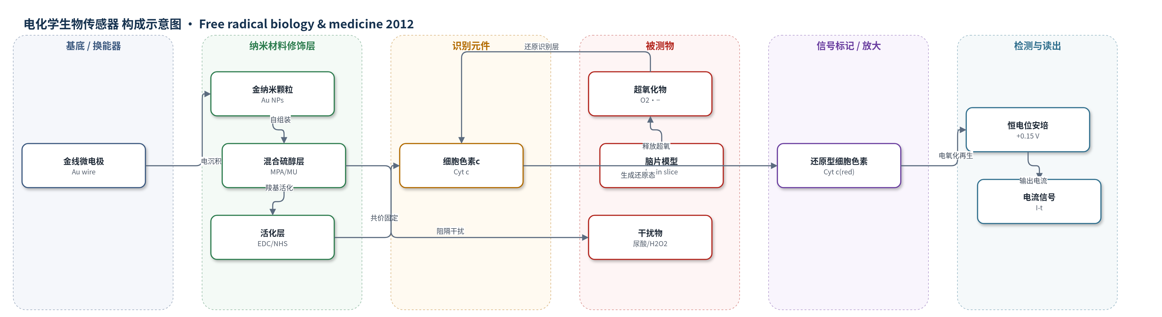
点击图片查看大图 · 依据论文自动绘制
传感器类型
电化学生物传感器
检测对象
超氧化物(superoxide anion radical, O2•−);样品基质:小鼠海马脑片(hippocampal brain slices)及人工脑脊液/培养液(culture medium),校准用PBS
检测原理
脑片释放的超氧化物(O2•−)扩散至金线微电极表面,与通过混合硫醇自组装层共价固定的细胞色素c(Cyt c)发生氧化还原反应,将Cyt c由氧化态还原为还原态。工作电极恒置于+0.15 V(vs Ag/AgCl),还原型Cyt c在电极界面被电氧化再生,形成与超氧稳态浓度相关的法拉第电流。校准采用HX/XOD体系生成超氧,并考虑其生成与歧化平衡;混合硫醇SAM和低电位阻隔尿酸、H2O2等干扰。外源SOD或纳米氧化铈清除超氧后电流下降,从而定量抗氧化活性。
检测灵敏度
LOD: 4.1 nM (PBS)、2.3 nM (culture medium);线性范围: 0–1.23 mM (PBS)、0–1.32 mM (culture medium);灵敏度: 11.78×10^2 A M−1 m−2 (PBS)、14.45×10^2 A M−1 m−2 (culture medium)
效应效果
传感器对尿酸(≤50 mM)和H2O2无响应,10 U/ml SOD可完全抑制1.04 mM超氧信号;脑片中每次加入500 U/ml SOD使电流下降约14%,三次后接近基线。传感器在PBS中4℃保存7天响应不变,循环伏安200次稳定。响应时间<1 s,4–5 s达稳态。灵敏度高于长链硫醇Cyt c电极(2.76×10^2 A M−1 m−2)和短链硫醇电极(0.56×10^2 A M−1 m−2)。脑片正常超氧1.95 mM,缺血升至4.1 mM;纳米氧化铈1 mg/ml每次降低14.5±4.5%(正常)或8.1±2.2%(缺血),相当于527 U SOD。
传感器的构成
- 工作电极基底:金线微电极(Au wire microelectrode,0.5 mm,尖端0.25 mm,L形)提供导电基底与信号转导。
- 纳米修饰层:电沉积金纳米颗粒(Au NPs,HAuCl4 0.01 M)增加表面粗糙度并促进Cyt c固定。
- 自组装单层:混合硫醇(MPA 1.25 mM、MU 3.75 mM)形成SAM,提供羧基并阻隔干扰物。
- 化学活化层:EDC/NHS(200 mM EDC、50 mM NHS)活化MPA羧基用于共价偶联。
- 识别元件:细胞色素c(Cyt c,5 μM)共价固定,作为超氧化物还原的电子介体。
- 辅助电极:Ag/AgCl/3 M NaCl参比电极与Pt丝对电极构成三电极体系。
- 储存介质:0.1 M PBS含100 mM EDTA(pH 7.5)清洗并4℃保存,维持Cyt c活性。
中文摘要
活性氧(ROS)过量产生及其损伤是多种疾病病理的核心。由于缺乏可连续测量的特异性探针和技术,ROS时空积累研究受限。我们展示了微型化电化学细胞色素c(Cyt c)生物传感器在小鼠急性脑片中实时测量天然及工程化抗氧化剂对超氧化物产生与清除的作用。对照条件下,400 μm厚脑片海马区产生的超氧化物处于电极检测范围内。缺血处理使超氧化物产生增加约两倍,信号在3–4 h内稳定。阴离子通道抑制剂DIDS显著降低对照条件下的胞外超氧化物信号,提示超氧化物经膜转运进入胞外空间可能参与正常氧化还原信号。外源超氧化物歧化酶(SOD)使信号呈剂量依赖性下降,验证电极对海马细胞释放超氧化物的特异性。平均直径15 nm的氧化铈纳米颗粒(nanoceria)清除超氧阴离子自由基活性相当于每1 mg/ml添加527 U SOD。本研究证明电化学生物传感器可用于研究生物模型中活性氧实时动态,并有助于界定超氧化物对氧化损伤的相对贡献。
英文摘要
The overproduction of reactive oxygen species and the resulting damage are central to the pathology of many diseases. The study of the temporal and spatial accumulation of reactive oxygen species has been limited because of the lack of specific probes and techniques capable of continuous measurement. We demonstrate the use of a miniaturized electrochemical cytochrome c (Cyt c) biosensor for real-time measurements and quantitative assessment of superoxide production and inactivation by natural and engineered antioxidants in acutely prepared brain slices from mice. Under control conditions, superoxide radicals produced from the hippocampal region of the brain in 400-μm-thick sections were well within the range of detection of the electrode. Exposure of the slices to ischemic conditions increased the superoxide production twofold and measurements from the slices were stable over a 3- to 4-h period. The stilbene derivative and anion channel inhibitor 4,4'-diisothiocyano-2,2'-disulfonic stilbene markedly reduced the extracellular superoxide signal under control conditions, suggesting that a transmembrane flux of superoxide into the extracellular space may occur as part of normal redox signaling. The specificity of the electrode for superoxide released by cells in the hippocampus was verified by the exogenous addition of superoxide dismutase (SOD), which decreased the superoxide signal in a dose-dependent manner. Similar results were seen with the addition of the SOD mimetic cerium oxide nanoparticles (nanoceria), in that the superoxide anion radical scavenging activity of nanoceria with an average diameter of 15 nm was equivalent to 527 U of SOD for each 1 μg/ml of nanoceria added. This study demonstrates the potential of electrochemical biosensors for studying real-time dynamics of reactive oxygen species in a biological model and the utility of these measurements in defining the relative contribution of superoxide to oxidative injury.