组成图示
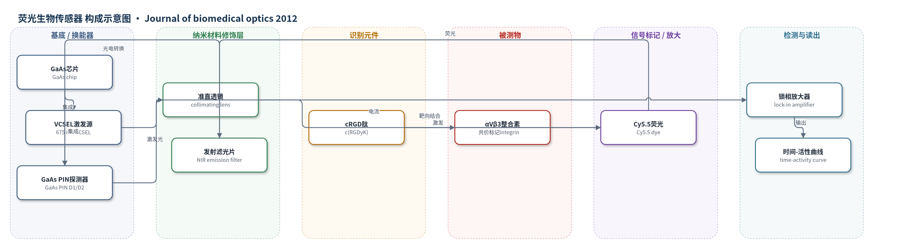
点击图片查看大图 · 依据论文自动绘制
传感器类型
荧光生物传感器
检测对象
cRGD-Cy5.5(靶向 αVβ3 整合素的近红外荧光分子探针)、cRAD-Cy5.5(非靶向对照探针)、Cy5.5(荧光染料);样品基质:荷 U87MG 胶质母细胞瘤小鼠的皮下肿瘤及对照组织(活体组织/血液分布)
检测原理
该传感器以 675 nm VCSEL 作为近红外激发源,激发光经准直透镜照射组织。静脉注射的 cRGD-Cy5.5 中,cRGD 环肽与肿瘤新生血管及肿瘤细胞表面高表达的 αVβ3 整合素特异性结合,使 Cy5.5 荧光染料在肿瘤区富集。Cy5.5 吸收激发光后发射近红外荧光,荧光经同一透镜收集,由探测器上方的近红外发射滤光片选择,再由 GaAs PIN 光电二极管转换为电流。锁相放大器读取 D1/D2 电流并扣除背景,得到时间-活性曲线。肿瘤区探针浓度越高,荧光越强,电流越大;通过双传感器肿瘤侧/对照侧比值及非靶向 cRAD-Cy5.5 校准,可定量肿瘤/背景比和动力学差异。
检测灵敏度
LOD: 5 nM in vitro;in vivo: autofluorescence-limited to 50 nM;in vitro tested concentrations: 250, 500, 2500, 10,000 nM (linear signal)
效应效果
传感器在开放、体模和活鼠条件下精确,CV% 0.291%–0.941%(N=20);长期稳定性 20–60 min,CV% <3.31%。重现性体模 9.7%–23.87%,活鼠 7.28%–14.07%。距离、角度和旋转改变信号幅度(0–15° 使 S1D1/S1D2 变化 112%/181%),但精度基本不变。双探测器在麻醉扰动下保持共线性,斜率约 0.979–0.989。与冷却 CCD 相机相比,cRGD-Cy5.5 校准后肿瘤/背景比 1 h 2.43±0.95、2 h 3.64±1.38;非靶向 cRAD-Cy5.5 比值约 0.476–0.542,靶向 cRGD-Cy5.5 比值 1.236–1.662。作者认为可用于连续监测分子探针动力学、肿瘤异质性和临床分子传感。
传感器的构成
- 基底/换能器芯片:GaAs 半导体芯片,集成 VCSEL 与 GaAs PIN 探测器,提供光电转换基底
- 激发光源:675 nm VCSEL 激光器,输出约 0.75–1.0 mW 近红外激发光
- 光学准直/封装:准直透镜与金属封装,准直激发光、收集荧光并固定器件
- 光电探测器:两个 GaAs PIN 光电二极管(D1/D2),将 Cy5.5 荧光转换为 pA 级电流
- 发射滤光片:集成近红外荧光发射滤光片(stacked filters),置于探测器上方,选择荧光并抑制激发泄漏
- 识别元件:cRGD 环肽(c(RGDyK)),作为分子探针靶向 αVβ3 整合素
- 信号标记物:Cy5.5 近红外荧光染料,标记 cRGD/cRAD 肽,被 VCSEL 激发后产生荧光信号
- 信号读出:锁相放大器、自动开关与激光驱动器,读取 D1/D2 电流并输出时间-活性曲线
中文摘要
分子光学成像是研究活体分子事件的重要技术,但现有方法难以在清醒、活动受试者中进行长期连续测量。为此,作者设计了一种新型轻量微型生物传感器,用于体内连续光学传感。该传感器包含封闭的垂直腔面发射半导体激光器(VCSEL)和相邻一对近红外光学滤波探测器,并采用双传感器同时探测正常与病变肿瘤位点。在体模和体内研究确认传感器精确后,作者在荷有人胶质母细胞瘤细胞的小鼠中使用靶向分子探针 cRGD-Cy5.5,该探针靶向肿瘤新生血管和肿瘤细胞表面的 αVβ3 整合素。传感器可捕获靶向分子探针的动态时间-活性曲线。cRGD-Cy5.5 注射后,经信号校准的平均肿瘤/背景比在 1 h 约为 2.43±0.95,2 h 约为 3.64±1.38(N=5),与冷却 CCD 相机获得的数据一致。作者认为该新型便携、精确生物传感器可用于多种疾病应用中分子探针的动力学和稳态水平评估。
英文摘要
Molecular optical imaging is a widespread technique for interrogating molecular events in living subjects. However, current approaches preclude long-term, continuous measurements in awake, mobile subjects, a strategy crucial in several medical conditions. Consequently, we designed a novel, lightweight miniature biosensor for in vivo continuous optical sensing. The biosensor contains an enclosed vertical-cavity surface-emitting semiconductor laser and an adjacent pair of near-infrared optically filtered detectors. We employed two sensors (dual sensing) to simultaneously interrogate normal and diseased tumor sites. Having established the sensors are precise with phantom and in vivo studies, we performed dual, continuous sensing in tumor (human glioblastoma cells) bearing mice using the targeted molecular probe cRGD-Cy5.5, which targets αVβ3 cell surface integrins in both tumor neovasculature and tumor. The sensors capture the dynamic time-activity curve of the targeted molecular probe. The average tumor to background ratio after signal calibration for cRGD-Cy5.5 injection is approximately 2.43±0.95 at 1 h and 3.64±1.38 at 2 h (N=5 mice), consistent with data obtained with a cooled charge coupled device camera. We conclude that our novel, portable, precise biosensor can be used to evaluate both kinetics and steady state levels of molecular probes in various disease applications.