组成图示
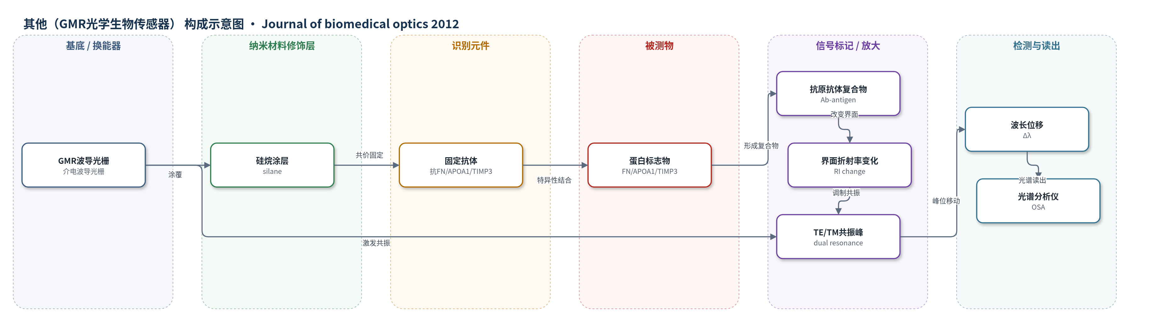
点击图片查看大图 · 依据论文自动绘制
传感器类型
其他(GMR光学生物传感器)
检测对象
纤连蛋白(fibronectin, FN)、载脂蛋白A1(apolipoprotein A1, APOA1)、基质金属蛋白酶组织抑制剂3(tissue inhibitor of metalloproteinases 3, TIMP3)、钙网蛋白(calreticulin)、I型胶原(collagen type 1)、丝裂原活化蛋白激酶13(mitogen-activated protein kinase 13, MAPK13);样品基质为卵巢癌细胞培养上清与培养基对照(非患者血清/血浆)
检测原理
GMR传感器板为亚波长介电波导光栅,宽带光入射时在表面激发TE和TM共振峰。传感器表面经硅烷修饰并用交联剂固定特异性抗体,BSA封闭后,样品中的目标蛋白与抗体结合。结合事件增加传感表面的光学厚度/界面折射率,使共振峰发生波长位移Δλ。光学光谱分析仪实时读取反射光谱,无需荧光或酶标记。信号大小与结合蛋白质量/浓度相关,通过重组蛋白标准曲线定量;TE/TM双峰交叉参考提高准确性。
检测灵敏度
TIMP3:免疫印迹检测限约2 μg/mL;RSI系统可常规测得低至0.05 μg/mL,最高2.61 μg/mL。MAPK13:理论检测阈值>1 μg/mL。纤连蛋白免疫印迹标准曲线:0.5–5 ng,R^2=0.987。GMR与免疫印迹相关:绝对浓度R^2=0.979,相对水平R^2=0.837;单项绝对R^2:FN 0.907、calreticulin 0.952、collagen type 1 0.573、APOA1 0.939;相对R^2:FN 0.932、calreticulin 0.868、collagen type 1 0.421、APOA1 0.992。
效应效果
GMR系统与定量免疫印迹在五种卵巢癌细胞系中高度一致,绝对浓度R^2=0.979,相对水平R^2=0.837。对TIMP3,免疫印迹未检出,GMR稳定测得0.05–2.61 μg/mL,TOV-21G和TOV-112D相对水平达5.80和5.00。纤连蛋白在Caov-3和SK-OV-3上清相对水平约2.06/2.07(免疫印迹)和2.27/1.86(GMR);载脂蛋白A1在TOV-112D升高、SK-OV-3降低。样品经BSA封闭和加标回收评估基质干扰,四重复平均。作者认为该系统适合卵巢癌蛋白标志物面板,尚需患者样本验证。
传感器的构成
- 基底/换能器:GMR传感器板(sub-wavelength dielectric waveguide grating,介电波导光栅),产生TE/TM共振峰并响应表面结合事件
- 表面化学修饰层:商用硅烷(silane)涂层,提供共价键合抗体的表面位点
- 识别元件:特异性抗体(anti-fibronectin MAB1918、anti-apolipoprotein A1、anti-calreticulin、anti-collagen type 1、anti-MAPK13、anti-TIMP3 MAB973),经交联剂(crosslinking agent)固定,捕获目标蛋白
- 封闭层:牛血清白蛋白(BSA)溶液,降低非特异性结合
- 标准/样品介质:PBS含3% BSA试剂稀释液,用于标准曲线、基线和加标回收;细胞上清/培养基直接孵育
- 光学读出:宽带光源与光学光谱分析仪(optical spectrum analyzer),实时监测共振波长位移Δλ
中文摘要
卵巢癌是致死率最高的妇科肿瘤,主要因诊断偏晚,目前不足20%的病例在早期被发现。可靠的早期诊断和疾病监测工具仍然缺乏,能够区分卵巢浆液性癌早晚期阶段的蛋白生物标志物尤为迫切。本研究在五种已建立的卵巢癌细胞系中,比较六种卵巢癌蛋白生物标志物的绝对和相对水平,采用定量免疫印迹和基于引导模共振(GMR)的无标记光学生物传感器检测系统进行验证。结果显示,GMR传感器能够准确、一致且可重复地定量蛋白标志物,并与定量免疫印迹结果高度相关,同时具有更高的灵敏度,适合用于新型卵巢癌生物标志物的定量与检测。研究进一步鉴定纤连蛋白、载脂蛋白A1和TIMP3可作为原发与转移性卵巢癌鉴别诊断的潜在蛋白标志物。未来仍需患者样本验证。
英文摘要
Ovarian carcinoma has the highest lethality rate of gynecologic tumors, largely attributed to the late-stage diagnosis of the disease. Reliable tools for both accurate diagnosis and early detection of disease onset are lacking, and presently less than 20% of ovarian cancers are detected at an early stage. Protein biomarkers that allow the discrimination of early and late stages of ovarian serous carcinomas are urgently needed as they would enable monitoring pre-symptomatic aspects of the disease, disease progression, and the efficacy of intervention therapies. We compare the absolute and relative protein levels of six protein biomarkers for ovarian cancer in five different established ovarian cancer cell lines, utilizing both quantitative immunoblot analysis and a guided-mode resonance (GMR) bioassay detection system that utilizes a label-free optical biosensor readout. The GMR sensor approach provided highly accurate, consistent, and reproducible quantification of protein biomarkers as validated by quantitative immunoblotting, as well as enhanced sensitivity, and is therefore suitable for quantification and detection of novel biomarkers for ovarian cancer. We identified fibronectin, apolipoprotein A1, and TIMP3 as potential protein biomarkers for the differential diagnosis of primary versus metastatic ovarian carcinoma. Future studies are needed to confirm the suitability of protein biomarkers tested herein in patient samples.