组成图示
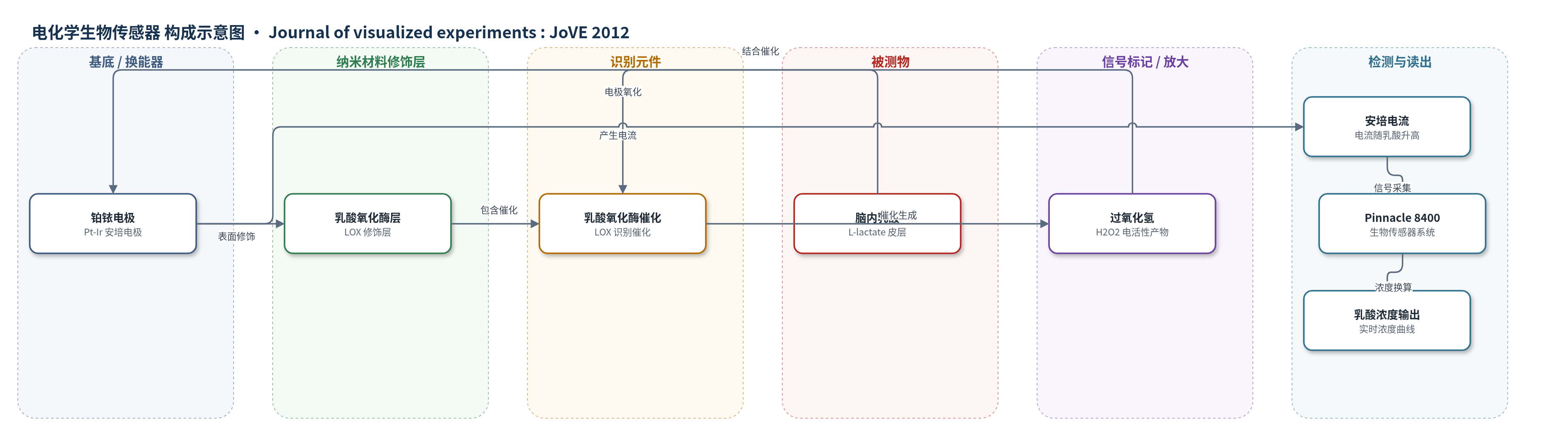
点击图片查看大图 · 依据论文自动绘制
传感器类型
电化学生物传感器
检测对象
乳酸(lactate, L-lactate);样品基质:小鼠额叶皮层脑组织(活体皮层)
检测原理
传感器探头植入小鼠额叶皮层,皮层乳酸扩散至铂铱电极表面的乳酸氧化酶(LOX)层。LOX 特异性催化乳酸氧化,反应生成过氧化氢(H2O2)。H2O2 在铂铱电极工作电位下发生安培氧化,产生与 H2O2 浓度相关的法拉第电流。当脑内糖酵解增强时,乳酸浓度升高,LOX 催化生成更多 H2O2,电极电流随之增大;当 NREMS 高幅慢波期间糖酵解降低时,乳酸和电流下降。系统实时采集电流并换算为乳酸浓度。该方法未使用 HCR、RCA 或 CRISPR-Cas 等额外放大策略,主要依赖酶催化与安培换能实现实时检测。
检测灵敏度
—
效应效果
该方法在自由行为小鼠中实现 EEG、EMG 与皮层乳酸的连续同步记录。乳酸传感器电流随睡眠状态变化:低幅 EEG 的清醒和 REMS 期升高,高幅 EEG 的 NREMS 期降低,表明可捕捉秒级糖酵解动态。光遗传刺激 1 Hz 或 10 Hz 时,EEG 双通道均响应;80–120 μW 通常使刺激频率 EEG 振幅增加约 50%,且远端记录支持波传播而非传感器光伪迹。传感器经体外乳酸标准液预校准。作者称该系统可用于研究自由行为啮齿动物中 EEG 水平神经元活动与脑内细胞能量关系,并兼容葡萄糖、谷氨酸等传感器。未报告 RSD、回收率或方法学对比数值。
传感器的构成
- 换能器电极:铂铱(Pt-Ir)电极,作为安培检测电极,响应过氧化氢氧化还原并产生电流
- 识别/催化元件:乳酸氧化酶(lactate oxidase, LOX)层,覆盖于电极表面,特异性催化乳酸氧化
- 信号产物:过氧化氢(H2O2),由 LOX 催化乳酸生成,在电极发生电化学反应形成电流
- 植入定位:颅骨固定乳酸导向套管(lactate guide cannula),用于将传感器探头置入额叶皮层
- 信号读出:Pinnacle 8400 生物传感器系统/前置放大器,采集电极电流并换算乳酸浓度
中文摘要
尽管脑组织质量不足体重的5%,静息时却消耗全身约四分之一的葡萄糖。非快速眼动睡眠(NREMS)占睡眠大部分时间,其功能尚不明确,但显著特征是脑葡萄糖利用较清醒时降低。NREMS 与脑电图中低于4 Hz的慢波相关,慢波反映皮层神经元在去极化上状态与超极化下状态间振荡;下状态期间神经元可数百毫秒不产生动作电位,而动作电位后离子梯度恢复是重要代谢负担,因此下状态可能降低代谢。为检验该假设,需要以秒级时间分辨率测量脑糖酵解代谢。作者通过检测有氧糖酵解产物乳酸来反映葡萄糖代谢速率,使用嵌入额叶皮层的乳酸氧化酶实时传感器。传感机制为铂铱电极外包裹乳酸氧化酶层,乳酸被氧化酶代谢生成过氧化氢,进而在铂铱电极产生电流;脑糖酵解增强时乳酸浓度升高,电极电流随之增大。同时,为分离皮层兴奋性,作者建立可同时记录脑电、乳酸生物传感器糖酵解通量,并光遗传激活锥体神经元操纵皮层活动的系统。该系统用于记录睡眠相关脑电波形与皮层乳酸浓度动态关系,适用于自由行为啮齿动物脑电与脑内能量研究。
英文摘要
Although the brain represents less than 5% of the body by mass, it utilizes approximately one quarter of the glucose used by the body at rest(1). The function of non rapid eye movement sleep (NREMS), the largest portion of sleep by time, is uncertain. However, one salient feature of NREMS is a significant reduction in the rate of cerebral glucose utilization relative to wakefulness(2-4). This and other findings have led to the widely held belief that sleep serves a function related to cerebral metabolism. Yet, the mechanisms underlying the reduction in cerebral glucose metabolism during NREMS remain to be elucidated. One phenomenon associated with NREMS that might impact cerebral metabolic rate is the occurrence of slow waves, oscillations at frequencies less than 4 Hz, in the electroencephalogram(5,6). These slow waves detected at the level of the skull or cerebral cortical surface reflect the oscillations of underlying neurons between a depolarized/up state and a hyperpolarized/down state(7). During the down state, cells do not undergo action potentials for intervals of up to several hundred milliseconds. Restoration of ionic concentration gradients subsequent to action potentials represents a significant metabolic load on the cell(8); absence of action potentials during down states associated with NREMS may contribute to reduced metabolism relative to wake. Two technical challenges had to be addressed in order for this hypothetical relationship to be tested. First, it was necessary to measure cerebral glycolytic metabolism with a temporal resolution reflective of the dynamics of the cerebral EEG (that is, over seconds rather than minutes). To do so, we measured the concentration of lactate, the product of aerobic glycolysis, and therefore a readout of the rate of glucose metabolism in the brains of mice. Lactate was measured using a lactate oxidase based real time sensor embedded in the frontal cortex. The sensing mechanism consists of a platinum-iridium electrode surrounded by a layer of lactate oxidase molecules. Metabolism of lactate by lactate oxidase produces hydrogen peroxide, which produces a current in the platinum-iridium electrode. So a ramping up of cerebral glycolysis provides an increase in the concentration of substrate for lactate oxidase, which then is reflected in increased current at the sensing electrode. It was additionally necessary to measure these variables while manipulating the excitability of the cerebral cortex, in order to isolate this variable from other facets of NREMS. We devised an experimental system for simultaneous measurement of neuronal activity via the elecetroencephalogram, measurement of glycolytic flux via a lactate biosensor, and manipulation of cerebral cortical neuronal activity via optogenetic activation of pyramidal neurons. We have utilized this system to document the relationship between sleep-related electroencephalographic waveforms and the moment-to-moment dynamics of lactate concentration in the cerebral cortex. The protocol may be useful for any individual interested in studying, in freely behaving rodents, the relationship between neuronal activity measured at the electroencephalographic level and cellular energetics within the brain.