组成图示
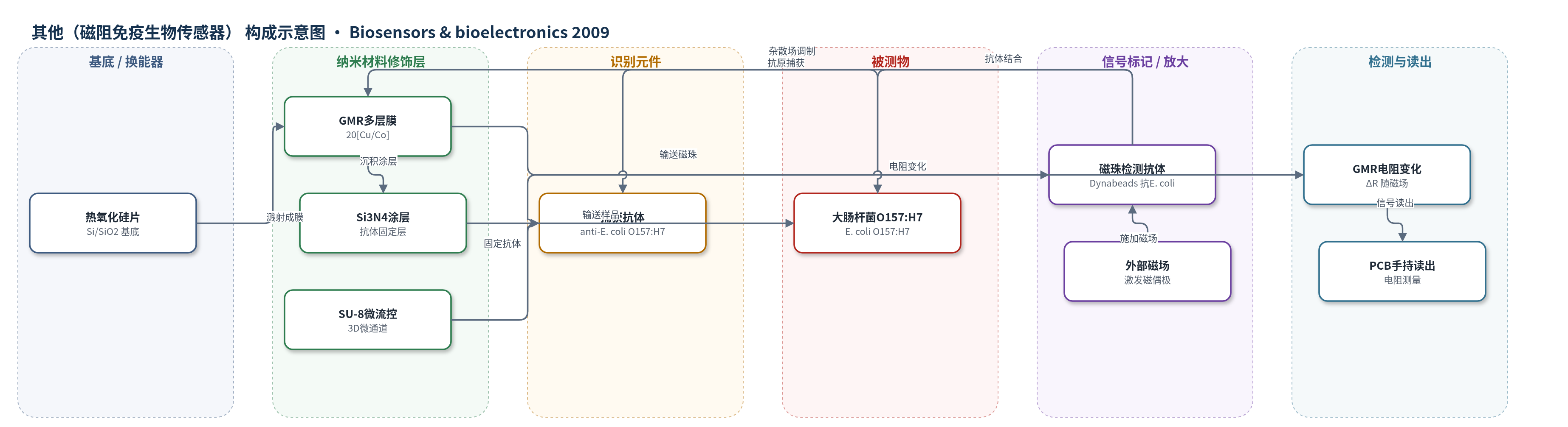
点击图片查看大图 · 依据论文自动绘制
传感器类型
其他(磁阻免疫生物传感器)
检测对象
大肠杆菌O157:H7(Escherichia coli O157:H7),样品基质为食品与临床样品
检测原理
样品经 SU-8 微流控网络进入传感区,固定在 Si3N4 表面的抗 E. coli O157:H7 抗体捕获目标菌。随后,抗 E. coli O157:H7 抗体包被的超顺磁微球流过,与菌体表面抗原结合,形成抗体—抗原—抗体夹心结构。施加外部磁场后,磁珠被磁化并产生杂散磁场,改变 GMR 多层膜中 Cu/Co 界面自旋相关散射和反铁磁耦合状态,使 GMR 电阻发生可测变化。磁珠数量随目标菌浓度增加,电阻变化相应增大,通过 PCB 读取电阻即可实现免疫磁定量。四通道布局和参考通道用于提高冗余与抗干扰。
检测灵敏度
灵敏度: 0.06 Ω/Oe(150–230 Oe);磁阻: 3.20% @235 Oe
效应效果
抗体特异性测试显示,对 E. coli O157:H7 在 10^5 CFU/ml 具有较高特异性,非致病 E. coli 仅在 10^7 CFU/ml 高浓度下检出,沙门氏菌阴性对照未检出。Si3N4 表面抗体吸附量高于 SU-8 和 SiO2,选作固定层。微流控通道在 0–100 kPa 下密封良好,实验流量与理论一致;50 kPa 染料测试表明各通道流速一致且无泄漏。GMR 退火后磁阻由 1.35% 提高到 3.20%(235 Oe),去磁后电阻可恢复初始值;矩形 meander 的磁阻和灵敏度约为纵向设计的 3.5 倍。作者预期磁珠在约 200 Oe 下可产生约 0.36% 的最小电阻变化,并认为手持封装便于样品更换和现场检测。
传感器的构成
- 基底/换能器电极:热氧化硅片(thermally oxidized silicon wafer,4 in,450 μm)承载器件,Pt 接触用于引出 GMR 电极。
- GMR 换能层:20[Cu5.10 nm/Co2.47 nm] 巨磁阻多层膜,经 300 ℃退火,235 Oe 磁阻 3.20%,检测磁珠杂散磁场引起的电阻变化。
- 表面涂层:PECVD 沉积 Si3N4 层,作为抗体固定表面,ELISA 显示抗体吸附高于 SU-8 和 SiO2。
- 识别元件:抗 E. coli O157:H7 抗体(Chemicon)固定于 Si3N4 表面,捕获样品中的目标菌。
- 信号标记物:抗 E. coli O157:H7 抗体包被超顺磁微球(Dynabeads,Dynal),与捕获菌形成夹心并提供磁偶极信号。
- 微流控层:SU-8 50/SU-8 5 光刻胶 3D 微通道与储液腔,两入口两出口,引导样品和磁珠至传感区并含参考通道。
- 封装/读出接口:立体光刻塑料上下胶囊、O-ring、PCB 金属引脚与标准软管,连接 GMR 电极和外部磁场/电阻测量系统。
中文摘要
本文报道了一种用于在食品和临床样品中免疫磁检测并定量致病性大肠杆菌O157:H7的手持式微系统。作者开发了将三维微流控网络与微加工生物传感器集成的芯片技术,完成了传感膜优化、微流控电路设计、生物检测协议开发以及安全便捷封装。该生物传感器通过抗体—抗原反应将抗原预先固定在传感表面,并检测与抗原结合的超顺磁微球引起的小磁场变化。传感膜采用巨磁阻多层结构,组成为20[Cu5.10 nm/Co2.47 nm],在235 Oe下磁阻为3.20%,在150–230 Oe范围内灵敏度可达0.06 Ω/Oe。由于硅氮化物适合抗体固定,被选为最佳传感表面涂层。SU-8光刻胶微流控网络用于将生物样品引导至传感区,立体光刻封装通过标准软管连接微通道,便于更换使用器件。
英文摘要
A hand held device has been designed for the immunomagnetic detection and quantification of the pathogen Escherichia coli O157:H7 in food and clinical samples. In this work, a technology to manufacture a Lab on a Chip that integrates a 3D microfluidic network with a microfabricated biosensor has been developed. With this aim, the sensing film optimization, the design of the microfluidic circuitry, the development of the biological protocols involved in the measurements and, finally, the packaging needed to carry out the assays in a safe and straightforward way have been completed. The biosensor is designed to be capable to detect and quantify small magnetic field variations caused by the presence of superparamagnetic beads bound to the antigens previously immobilized on the sensor surface via an antibody-antigen reaction. The giant magnetoresistive multilayer structure implemented as sensing film consists of 20[Cu(5.10nm)/Co(2.47 nm)] with a magnetoresistance of 3.20% at 235Oe and a sensitivity up to 0.06 Omega/Oe between 150Oe and 230Oe. Silicon nitride has been selected as optimum sensor surface coating due to its suitability for antibody immobilization. In order to guide the biological samples towards the sensing area, a microfluidic network made of SU-8 photoresist has been included. Finally, a novel packaging design has been fabricated employing 3D stereolithographic techniques. The microchannels are connected to the outside using standard tubing. Hence, this packaging allows an easy replacement of the used devices.