组成图示
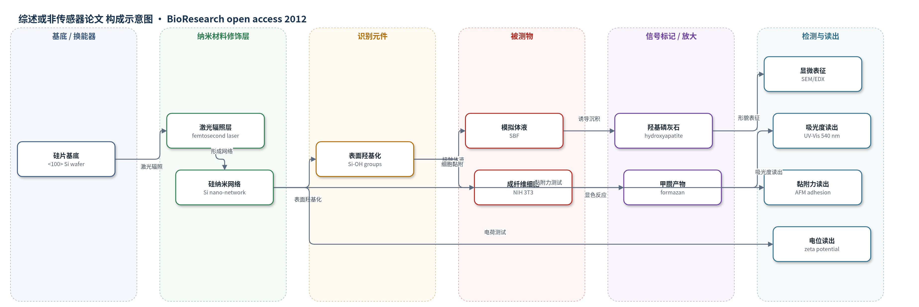
点击图片查看大图 · 依据论文自动绘制
传感器类型
综述或非传感器论文
检测对象
未报道明确检测对象(非传感检测论文);涉及样品基质:模拟体液(SBF)、细胞培养体系(NIH 3T3成纤维细胞)
检测原理
本文未报道生物传感器检测原理,而是研究硅纳米网络的生物活性机制。飞秒激光在硅片表面合成互连硅纳米网络,形成类似细胞外基质的三维多孔结构。该网络具有较高负电荷和表面Si-OH基团,可吸附蛋白并增强细胞黏附。在模拟体液中,Si-OH促进Ca2+和磷酸根离子成核,形成类骨羟基磷灰石;块体硅14天无沉积,而纳米网络3天即可沉积。NIH 3T3细胞在网络上铺展、迁移和增殖,MTT法通过甲臜吸光度反映细胞增殖。文中未涉及识别元件、信号放大或传感读出机制。
检测灵敏度
未报道(非传感器论文)
效应效果
与块体硅相比,硅纳米网络在模拟体液中3天即形成羟基磷灰石,10天后表面被磷灰石覆盖;块体硅14天无沉积。zeta电位较空白硅提高约1.6倍,100 μm厚度样品的AFM黏附力约为空白硅的7倍。NIH 3T3成纤维细胞增殖较块体硅提高约300%,t检验p=0.002,表明差异显著。SEM显示细胞具有多伪足、铺展和迁移形态。论文未报告选择性、抗干扰、稳定性、RSD、实际样品回收率或与ELISA/HPLC/qPCR等方法的定量对比,但作者认为该ECM样硅纳米网络可用于组织工程、再生医学及生物传感器检测等应用。
传感器的构成
- 基底:未掺杂<100>取向硅片(Si wafer,500 μm厚,4 cm²),作为支撑基底与硅源
- 纳米网络层:飞秒激光合成互连硅纳米纤维/纳米颗粒网络(Si nano-network),形成ECM样三维多孔结构
- 表面化学层:硅表面Si-OH基团(由SBF中水分子作用形成),促进蛋白吸附与羟基磷灰石成核
- 生物活性产物层:模拟体液孵育后沉积的类骨羟基磷灰石(hydroxyapatite, HA),体现生物活性
- 细胞相互作用层:NIH 3T3小鼠胚胎成纤维细胞(NIH 3T3 fibroblasts),用于评估细胞黏附、铺展与增殖
- 表征/读出层:SEM、TEM、EDX、zeta电位、AFM、MTT比色法,用于形貌、电荷、黏附力与细胞增殖分析
中文摘要
纳米结构硅在组织工程中具有应用前景,但现有硅支架制备多依赖昂贵、复杂且耗时的光刻技术,并需使用腐蚀性化学品,所得结构也难以真正模拟细胞外基质(ECM)。本文报道了一种由兆赫兹超快激光合成制备的互连硅纳米网络。该方法简单、快速、无需添加任何化学物质,并可在环境条件下完成。通过改变激光参数,可调节硅纤维网络的孔径和密度。显微分析表明,该硅网络具有高表面电荷和较强的黏附力。体外生物活性测试显示,其在模拟体液中仅3天即可沉积类骨羟基磷灰石。细胞增殖实验表明,与块体硅相比,硅纳米网络使NIH 3T3成纤维细胞增殖提高约300%。扫描电镜观察显示细胞在网络上健康迁移和黏附。研究提示细胞增殖增强与ECM样硅纳米网络结构相关,该材料在组织工程、再生医学及生物传感器检测等生物医学应用中具有潜力。
英文摘要
Nanostructured silicon has proven to be a promising candidate in tissue engineering. However, recent research on fabrication of silicon scaffolds has been limited to expensive, complex, and time-consuming lithographic techniques that require the addition of caustic chemicals. Moreover, these techniques generate structures that do not truly mimic the extracellular matrix (ECM). Therefore, we introduce a novel, interlinked, silicon nano-network fabricated by MHz ultrafast laser synthesis. We demonstrate that ultrafast laser synthesis is simple, rapid, free of any chemical additions, and can be carried out under ambient conditions. Variation in laser parameters resulted in an alteration in the pore size and density of the silicon fibrous network. Microscopic analysis revealed a highly charged silicon network with elevated adhesion forces. In vitro bioactivity tests indicate the precipitation of bone-like apatite in just 3 days. Cell proliferation studies on the silicon nano-network present a 300% increase in comparison to its bulk counterpart. Scanning electron microscopy analysis shows healthy migration and attachment of cells on the silicon nano-network. This study points to a correlation between elevated cell proliferation and the ECM-like structure of the silicon nano-network. This ECM-like silicon nano-network suggests significant potential not only in tissue engineering and regeneration but also in other biomedical applications such as biosensor detection.