组成图示
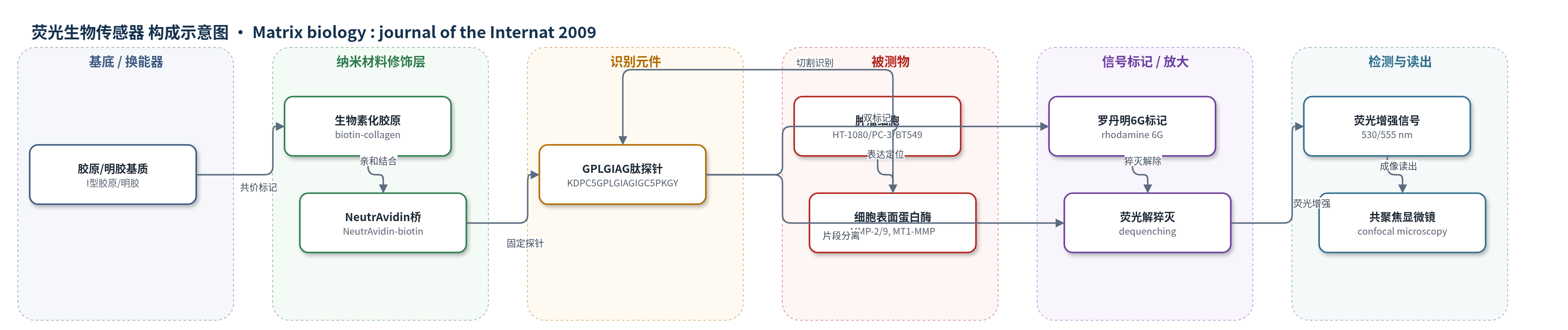
点击图片查看大图 · 依据论文自动绘制
传感器类型
荧光生物传感器
检测对象
蛋白酶活性(protease activity,主要为 MMP-2、MMP-9、MT1-MMP/MMP-14);样品基质:活肿瘤细胞迁移的二维明胶(gelatin)或三维 I 型胶原(type I collagen)基质
检测原理
该传感器以含间质胶原切割位点 GPLGIAG 的十八肽为识别元件,肽链两端以罗丹明6G双标记,形成分子内 H 型激基二聚体并发生荧光自猝灭。当肿瘤细胞表面或细胞外基质中的蛋白酶(如 MMP-2、MMP-9、MT1-MMP)识别并切割 GPLGIAG 位点时,肽链断裂为两个片段,一个片段仍固定在基质上,另一片段扩散离开,从而破坏罗丹明6G二聚体的分子内猝灭,产生局部红色荧光。荧光出现的位置和强度随局部蛋白酶活性变化,因此可实时成像蛋白酶活性位点。D-肽对照不被蛋白酶切割,用于排除胶原聚集、机械张力等物理效应造成的假信号。
检测灵敏度
—
效应效果
探针具立体特异性,L-肽可被多种蛋白酶切割,D-肽不切割;切割速率如 MMP-9 447.8 fmol/s、MMP-2 337.0 fmol/s、MT1-MMP 99.8 fmol/s。TIMP-2 抑制膜结合 MMP 97.1–99.9%、MMP-2 97.1%、MMP-9 86.0%;TIMP-1 抑制 MMP-2 98.3%、MMP-9 49.3%,不抑制膜结合 MMP。与 DQ-collagen 相比,探针在胶原聚集处背景更低。交联胶原中,TIMP-2 使 HT-1080 速度由 43.8±11.5 μm/h 降至 6.73±3.04 μm/h(85%,P<0.001),TIMP-1 降至 29.2±8.3 μm/h(约33%);胃蛋白酶胶原中无抑制。作者认为其可高分辨率定位活细胞蛋白酶活性。
传感器的构成
- 基底/基质:I型胶原(type I collagen)或明胶(gelatin)二维/三维基质,承载探针并提供细胞迁移微环境
- 识别元件:含 GPLGIAG 切割位点的十八肽(KDPC5GPLGIAGIGC5PKGY,C5=6-aminohexanoic acid),响应 MMP-2、MMP-9、MT1-MMP 等蛋白酶切割
- 信号标记物:罗丹明6G(rhodamine 6G)双标记,形成自猝灭 H 型激基二聚体,切割后解离产生荧光
- 固定/连接层:生物素(biotin)-NeutrAvidin 亲和连接,将探针固定于生物素化胶原/明胶基质
- 对照元件:D-肽对照(all d-amino acids)探针,排除基质聚集、张力等非特异物理效应
中文摘要
确定蛋白酶在细胞迁移和侵袭中的具体作用,需要在活细胞穿过细胞外基质时对其蛋白酶活性位点进行高分辨率成像。作者设计了一种新型荧光生物传感器,用于检测细胞外局部蛋白酶活性,并检验基质金属蛋白酶(MMP)功能在细胞迁移和侵袭三维胶原基质中的必要性。该探针含有间质胶原中的肽切割位点,可被 MMP-2、MMP-9 和 MMP-14(MT1-MMP)等多种蛋白酶切割后发出荧光,且无需转染或修饰被检测细胞。利用该生物传感器衍生化的基质,作者证明蛋白酶活性位于迁移肿瘤细胞极化的前缘,而不是细胞体后部。该蛋白酶活性对天然交联胶原基质中的细胞迁移必不可少,但对胃蛋白酶处理胶原基质则非必需。这种新型高分辨率探针可提供蛋白酶活性的位点特异性报告,有助于理解细胞穿过细胞外基质迁移的机制,并澄清既往关于蛋白酶在转移中作用的研究差异。
英文摘要
Determining the specific role(s) of proteases in cell migration and invasion will require high-resolution imaging of sites of protease activity during live-cell migration through extracellular matrices. We have designed a novel fluorescent biosensor to detect localized extracellular sites of protease activity and to test requirements for matrix metalloprotease (MMP) function as cells migrate and invade three-dimensional collagen matrices. This probe fluoresces after cleavage of a peptide site present in interstitial collagen by a variety of proteases including MMP-2, -9, and -14 (MT1-MMP) without requiring transfection or modification of the cells being characterized. Using matrices derivatized with this biosensor, we show that protease activity is localized at the polarized leading edge of migrating tumor cells rather than further back on the cell body. This protease activity is essential for cell migration in native cross-linked but not pepsin-treated collagen matrices. The new type of high-resolution probe described in this study provides site-specific reporting of protease activity and insights into mechanisms by which cells migrate through extracellular matrices; it also helps to clarify discrepancies between previous studies regarding the contributions of proteases to metastasis.