组成图示
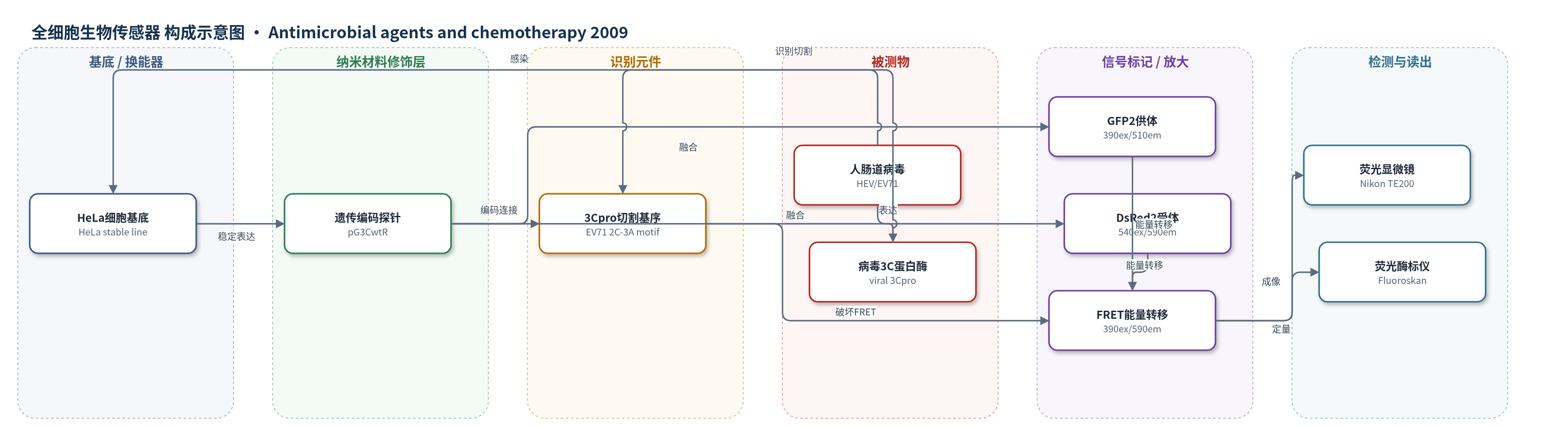
点击图片查看大图 · 依据论文自动绘制
传感器类型
全细胞生物传感器
检测对象
人肠道病毒(human enterovirus, HEV;含 EV71、CVA16、CVB1–CVB4、Echo9)感染及其 3C 蛋白酶活性;样品基质为 HeLa 细胞培养物(病毒感染细胞)
检测原理
该传感器基于遗传编码 FRET 探针。HeLa 细胞稳定表达 GFP2-3Cpro 切割基序-DsRed2 融合蛋白;未感染时,GFP2 与 DsRed2 距离在 1–10 nm 内,390 nm 激发 GFP2 后能量非辐射转移至 DsRed2,产生约 590 nm 红色发射,FRET 比率(590/510)较高。HEV 进入细胞后复制并表达 3C 蛋白酶,3Cpro 特异性切割 EV71 2C-3A 基序,使 GFP2 与 DsRed2 物理分离,FRET 效率下降,绿色 510 nm 发射相对增加、红色 590 nm 发射相对减少。病毒滴度越高、感染时间越长,3Cpro 产生越多,切割越充分,FRET 下降越大。rupintrivir 抑制 3Cpro 后切割减少,FRET 保持较高,从而可定量 EC50。
检测灵敏度
相关系数:R^2 = 0.985(FRET 与 PRA EC50);R^2 = 0.848(FRET 与 DRA EC50)
效应效果
该 FRET 生物传感器对 EV71、CVA16、CVB1–CVB4 和 Echo9 等 HEV 血清型均产生剂量依赖的 FRET 下降,而对 HSV-1 无响应;突变切割基序细胞也不响应,说明特异性来自 3Cpro 切割。Western blot 显示 EV71 感染后约 60 kDa 融合蛋白减少并出现约 30 kDa 切割产物。药敏实验中,FRET 法与 PRA 的 EC50 相关系数为 R^2=0.985,与 DRA 为 R^2=0.848;例如 EV71 的 rupintrivir EC50 分别为 0.781±0.028、0.791±0.167 和 0.561±0.083 μM。FRET 法仅需 36–48 h,短于 PRA 的 4–7 d 和 DRA 的 2–4 d,数据以三次重复均值±标准差表示,作者认为其可用于快速定量 HEV 感染和 3Cpro 抑制剂高通量筛选。
传感器的构成
- 细胞基底:HeLa 细胞,作为稳定表达 FRET 探针的活细胞平台,提供病毒吸附、感染与 3C 蛋白酶表达环境。
- 遗传编码探针:pG3CwtR 质粒编码的 GFP2-3Cpro 切割基序-DsRed2 融合蛋白,作为可被病毒蛋白酶切割的 FRET 报告分子。
- 识别元件:EV71 3Cpro 切割基序(2C-3A 连接区,IEALFQ-GPPKFR),作为人肠道病毒 3C 蛋白酶的特异性识别与切割位点。
- 荧光供体:GFP2,位于切割基序一侧,390 nm 激发后发射约 510 nm 绿光,并在未切割时向 DsRed2 转移能量。
- 荧光受体:DsRed2,位于切割基序另一侧,发射约 590 nm 红光,作为 FRET 受体。
- 对照探针:pG3CmutR 编码的突变切割基序(IEKLFQPPPKFR),用于验证 3Cpro 切割特异性。
- 信号读出:FRET 比率(590/510)荧光信号,经 Nikon TE200 荧光显微镜与 Fluoroskan Ascent 374 荧光酶标仪读取。
中文摘要
本研究建立了一种基于荧光共振能量转移(FRET)的实时检测系统,用于监测人肠道病毒(HEV)感染细胞内 3C 蛋白酶活性并评价抗病毒药物效力。作者通过遗传工程使 HeLa 细胞稳定表达一种 FRET 探针,该探针由绿色荧光蛋白 2(GFP2)和红色荧光蛋白 2(DsRed2)组成,两者之间连接 EV71 3C 蛋白酶切割基序。稳定细胞在未感染时呈现有效 FRET;HEV 感染后,病毒 3C 蛋白酶切割探针,使 FRET 被破坏,荧光发射由约 600 nm 红光向约 510 nm 绿光变化,可经荧光显微镜实时观察。荧光酶标仪定量显示,FRET 抑制程度与输入病毒滴度和感染时间呈剂量-时间依赖关系。该 FRET 生物传感器对多种相关 HEV 血清型有响应,但对系统发育距离较远的单纯疱疹病毒 1 型无响应,并经 Western blot 验证。进一步将 FRET 生物传感器用于 3C 蛋白酶抑制剂 rupintrivir 的药敏检测,结果显示其对代表性 HEV 血清型的敏感性测定准确。总之,该新型 FRET 系统可用于 HEV 的快速检测、定量和药物敏感性测试,并具有高通量筛选潜力。
英文摘要
A real-time assay system that allows monitoring of intracellular human enterovirus (HEV) protease activity was established using the principle of fluorescence resonance energy transfer (FRET). It was accomplished by engineering cells to constitutively express a genetically encoded FRET probe. The FRET-based probe was designed to contain an enterovirus 71 3C protease (3C(pro)) cleavage motif flanked by the FRET pair composed of green fluorescent protein 2 and red fluorescent protein 2 (DsRed2). Efficient FRET from the stable line was detected in a real-time manner by fluorescence microscopy, and the disruption of FRET was readily monitored upon HEV infection. The level of the repressed FRET was proportional to the input virus titer and the infection duration as measured by the fluorometric method. The FRET biosensor cell line was also responsive to other related HEV serotypes, but not to the phylogenetically distant herpes simplex virus, which was confirmed by Western blot analysis. The FRET biosensor was then utilized to develop a format for the determination of antiviral susceptibility, as the reduced FRET appeared to reflect viral replication. Evaluations of the FRET biosensor system with representative HEV serotypes demonstrated that their susceptibilities to a 3C(pro) inhibitor, rupintrivir, were all accurately determined. In summary, this novel FRET-based system is a means for rapid detection, quantification, and drug susceptibility testing for HEVs, with potential for the development of a high-throughput screening assay.