组成图示
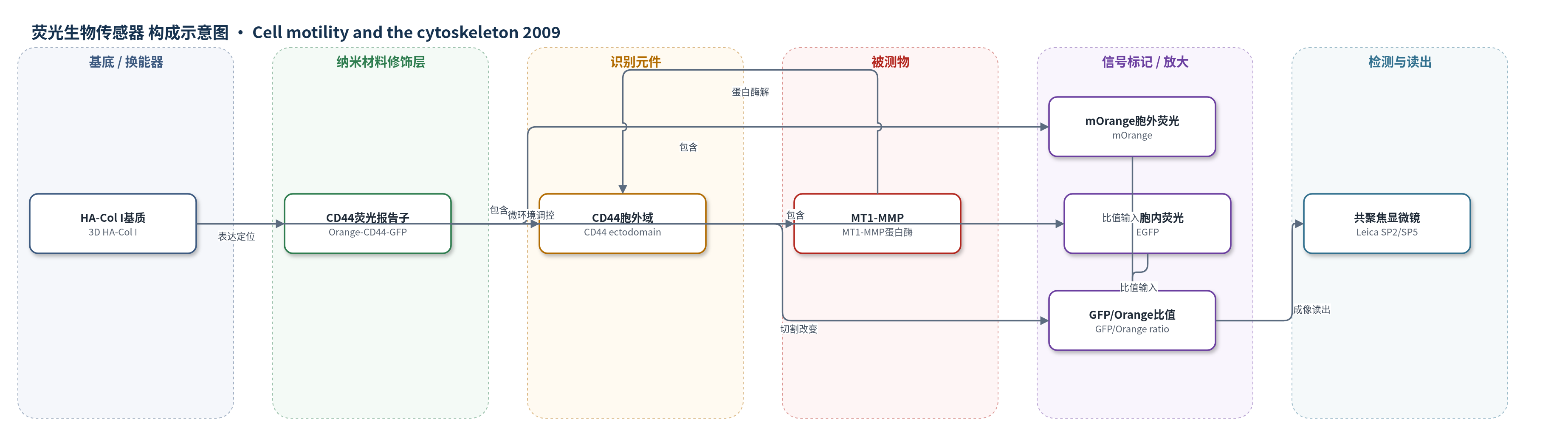
点击图片查看大图 · 依据论文自动绘制
传感器类型
荧光生物传感器
检测对象
CD44 胞外域切割/脱落(CD44 shedding/cleavage,MT1-MMP 介导);样品基质:活肿瘤细胞(MDA-MB-231、HT-1080、BLM)在 3D HA-Col I 基质或 HA 包被盖玻片上。
检测原理
该荧光生物传感器将 mOrange 融合于 CD44 胞外域、EGFP 融合于 CD44 胞内域,形成 Orange-CD44-GFP 报告子。当 MT1-MMP 在细胞膜上识别并切割 CD44 胞外域时,胞外 mOrange 标记片段从细胞表面释放,而胞内 EGFP 保留在细胞内,导致细胞内 EGFP 与胞外 mOrange 的荧光比值升高。串联 GFP-Orange 对照用于校正两种荧光蛋白量子产率差异。共聚焦显微镜采集 GFP 与 Orange 通道图像并生成比值图像,比值越高表示 CD44 切割/脱落越强。因此信号随 MT1-MMP 活性或 CD44 切割程度增加而增强,GM6001、抗 MT1-MMP 抗体或 siRNA 沉默可抑制该信号。
检测灵敏度
原文未报告 LOD、线性范围、灵敏度斜率或相关系数。
效应效果
该报告子可定量监测 CD44 切割:PMA 处理后 135 kD 全长 Orange-CD44-GFP 减少,52 kD 胞内 CD44-GFP 片段出现,GM6001 或 MT1-MMP siRNA 可抑制切割。在 HA-3D Col I 中,抗 MT1-MMP 抗体 Lem-2/15 完全消除 CD44 脱落,siRNA 亦降低切割。表达 Orange-CD44-GFP 使细胞侵袭增加 39.6%,且被 GM6001 抑制。CD44 过表达促进侵袭,阻断 CD44 抑制该效应;阻断 MT1-MMP 降低 MT1-MMP 与 CD44 介导的侵袭,而阻断 CD44 不影响 MT1-MMP 介导侵袭。BLM 细胞中 CD44-GFP 在 MT1-MMP 完整时增加侵袭 16%,在 MT1-MMP 沉默时抑制 74%。作者认为该工具可用于研究膜蛋白胞外域脱落及 MT1-MMP 在肿瘤侵袭中的作用。
传感器的构成
- 微环境/基底:3D HA-Col I 基质(HA 透明质酸 + Col I 胶原 I)及 HA 包被盖玻片,提供侵袭微环境并诱导 CD44 切割
- 识别元件:Orange-CD44-GFP 报告子中的 CD44 胞外域,作为 MT1-MMP 蛋白酶解底物/识别位点
- 信号标记物:mOrange 与 EGFP 荧光蛋白,分别融合于 CD44 胞外域和胞内域,切割后通过 GFP/Orange 比值报告
- 校准对照层:tandem GFP-Orange 串联荧光蛋白,用于校正 GFP 与 Orange 量子产率差异并提供 1:1 对照
- 读出系统:Leica SP2/SP5 共聚焦显微镜与 MetaMorph/Imaris 图像分析,采集 GFP/Orange 比值图像并定量
中文摘要
CD44 与膜型基质金属蛋白酶 MT1-MMP 在肿瘤细胞中协同促进侵袭,但机制不清。作者在含透明质酸(HA)的三维胶原 I(3D HA-Col I)基质中,利用活细胞时间序列共聚焦显微镜研究二者相互作用。荧光共振能量转移(FRET)显微成像显示,CD44 与 MT1-MMP 的相互作用优先发生在侵袭肿瘤细胞的后缘回缩部位以及侵袭过程中释放的膜片段上。作者进一步设计了一种用于活细胞成像的荧光生物传感器,以监测 CD44 的蛋白酶解加工,结果显示 CD44 胞外域切割富集于侵袭肿瘤细胞回缩的后端。侵袭实验表明,MT1-MMP 介导 CD44 依赖性肿瘤细胞侵袭,而 CD44 并非 MT1-MMP 介导 3D HA-Col I 基质侵袭所必需。结果支持 MT1-MMP 在 CD44 介导细胞侵袭的细胞回缩过程中发挥关键作用。
英文摘要
The adhesion molecule CD44 and the membrane-type matrix metalloproteinase MT1-MMP act coordinately in tumor cells to promote cell invasion through a yet unclear mechanism. We are interested in studying the interplay between CD44 and MT1-MMP in carcinoma cells embedded in HA containing three-dimensional collagen I matrices (3D HA-Col I) by time-lapse confocal microscopy imaging. Here we report the in vivo interaction between CD44 and MT1-MMP, revealed by fluorescence resonance energy transfer (FRET) microscopy. MT1-MMP interacts with CD44 preferentially at the trailing edge of the invading tumor cells during rear retraction and on membrane fragments released during the invasion process. A fluorescent biosensor designed to monitor the proteolytic processing of CD44 by live cell imaging demonstrates that cleavage of the CD44 extracellular domain is enriched in the retracting rear ends of invasive tumor cells. Invasion assays showed that MT1-MMP mediates CD44-dependent tumor-cell invasion, whereas CD44 is not essential for MT1-MMP-mediated invasion of 3D HA-Col I matrices. Together, our results support a role for MT1-MMP in cell retraction during CD44-mediated cell invasion.