组成图示
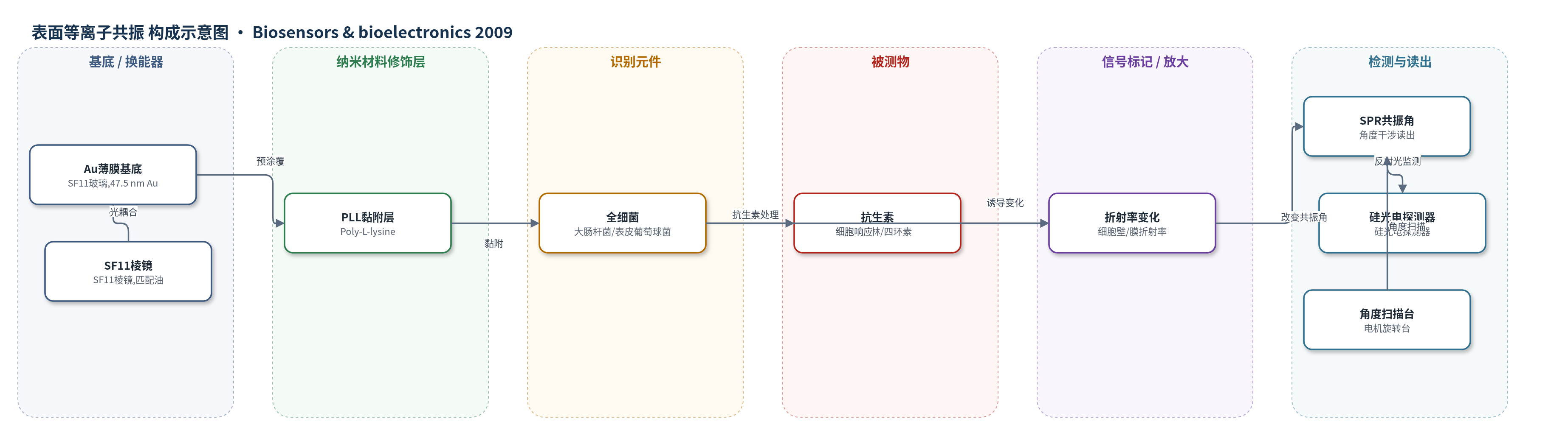
点击图片查看大图 · 依据论文自动绘制
传感器类型
表面等离子共振(SPR)生物传感器
检测对象
细菌抗菌药敏性(antimicrobial susceptibility):大肠杆菌 JM109(Escherichia coli JM109)对氨苄西林(ampicillin)、表皮葡萄球菌(Staphylococcus epidermidis)对四环素(tetracycline);样品基质为 LB 肉汤/去离子水流路中的细菌悬液及抗生素溶液。
检测原理
该传感器采用 Kretschmann 构型 SPR 角度干涉。细菌经 Poly-L-lysine 非特异性黏附于 Au 薄膜表面,形成全细胞识别层;随后流路注入抗生素。抗生素作用于细菌细胞壁、细胞膜或胞内蛋白合成等过程,使靠近 Au 界面约 100–300 nm 范围内的细菌结构松弛、破裂或含水量/折射率发生变化。由于 SPR 倏逝场对金属-介质界面附近折射率变化高度敏感,细菌折射率变化会改变表面等离子体激元的耦合效率,使共振角发生偏移。敏感菌因细胞壁/膜受损或生理状态改变,SPR 角偏移更大或呈不规则变化;耐药菌角偏移较小且较稳定。系统通过硅光电探测器实时监测反射光强度随入射角的变化,从而定性区分敏感株与耐药株。该方法不依赖细菌倍增时间,也不需特异性抗体或外源标记。
检测灵敏度
灵敏度: 约 1.4 × 10⁻⁴(折射率);对应 SPR 角偏移: 0.00867°
效应效果
该方法可在抗生素处理少于 2 h 内完成药敏判断,而常规 Kirby-Bauer 纸片扩散法和肉汤微量稀释法通常需要 1 天至数周。对 10 组大肠杆菌 JM109 的重复测试中,耐药株平均 SPR 角偏移为 −0.00527°,标准差 0.00763°;敏感株平均为 −0.01835°,标准差 0.00602°,敏感株判断正确率 100%,耐药株 80%,双尾 p 值为 4.78×10⁻⁴,具有统计学显著性。表皮葡萄球菌对四环素的盲测结果与已知药敏属性一致,且重复多次均观察到敏感株不规则角偏移。SEM 显示 30 min 内敏感株形态变化不明显,而 SPR 可检测到早期变化,说明其灵敏度高于 SEM。作者认为该方法可缩短临床等待时间,但当前结果偏定性,尚需建立定量药敏关系并开发多通道芯片。
传感器的构成
- 换能基底:SF11 玻璃基底溅射 47.5 nm Au 薄膜,作为 SPR 激发界面;SF11 棱镜与匹配油耦合激光
- 黏附修饰层:Poly-L-lysine(PLL)预涂覆于 Au 薄膜,提供细菌非特异性黏附
- 识别/响应元件:全细胞层,E. coli JM109 或 S. epidermidis 菌株黏附于 PLL/Au 表面,作为药敏检测对象
- 信号标记物:无外源荧光/酶标记,依赖细菌细胞壁/膜折射率变化产生 SPR 信号
- 流体模块:带流道的细胞腔室与 O-ring 密封,用于注入 LB 肉汤、去离子水和抗生素
- 温控模块:热敏电阻(thermistor)与 TE 制冷器维持 37°C,温度波动小于 0.1°C
- 光学读出:632.8 nm He-Ne 激光、硅光电探测器与角度扫描台,监测反射光强度并确定 SPR 共振角
中文摘要
本研究利用表面等离子共振(SPR)生物传感器的高灵敏度,探索细菌耐药性的快速检测方法。以大肠杆菌 JM109(Escherichia coli JM109)对氨苄西林(ampicillin)的敏感株和耐药株,以及表皮葡萄球菌(Staphylococcus epidermidis)对四环素(tetracycline)的敏感株和耐药株为对象,将细菌黏附于金薄膜表面,通过流路注入抗生素,实时记录细菌对抗生素响应引起的光学性质变化。结果显示,氨苄西林敏感株的 SPR 角偏移通常超过耐药株的 3 倍,四环素敏感株出现不规则 SPR 角偏移,而耐药株 SPR 角几乎不变。该 SPR 药敏方法抗生素处理时间少于 2 h,无需特异性抗体,相比常规 Kirby-Bauer 纸片扩散法和肉汤微量稀释法通常需 1 天至数周,可显著缩短实验室检测等待时间,有利于需要及时抗生素治疗的患者。所构建 SPR 生物传感器的折射率灵敏度约为 1.4×10⁻⁴。
英文摘要
Utilizing the ultra sensitivity of surface plasmon resonance (SPR) biosensor to examine drug resistance of bacteria was studied in this research. Susceptible and resistant strains of Escherichia coli JM109 to ampicillin and those of Staphylococcus epidermidis to tetracycline, served as a blind test, were examined. The bacteria adhered on the Au thin film was treated by the injection of antibiotic flow. The optical property change of the bacteria responded to antibiotics were recorded through SPR mechanism. As a result, the susceptible strain of E. coli generally revealed more than three times of SPR angle shift when compared to the resistant one; the susceptible strain of S. epidermidis revealed irregular SPR angle shift while the resistant strain kept the SPR angle almost unchanged. The new SPR method took less than 2h of antibiotic treatment time to complete the antimicrobial susceptibility test. Different from conventional applications of SPR, specific antibodies is not required in this method. As compared to the conventional assays, Kirby-Bauer disk diffusion and variations of broth microdilution usually take 1 day to weeks to issue the report. Using this SPR assay can greatly reduce the waiting period for laboratory tests, and can therefore benefit patients who need proper antibiotic treatments to control bacterial infections. The sensitivity of the SPR biosensor built for the application is around 1.4 x 10(-4) on the refractive index.