组成图示
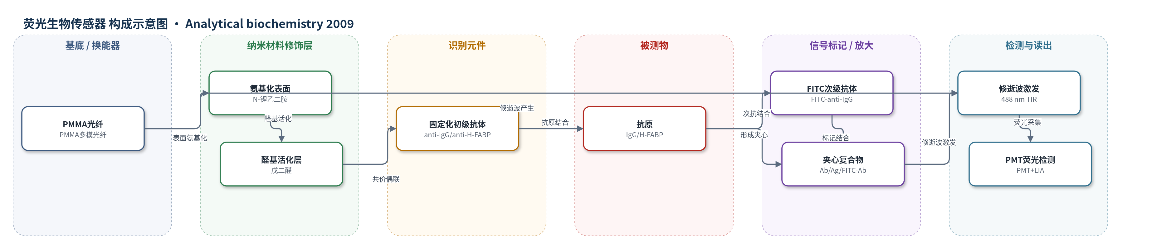
点击图片查看大图 · 依据论文自动绘制
传感器类型
荧光生物传感器
检测对象
小鼠免疫球蛋白G(mouse IgG)、人型脂肪酸结合蛋白(human H-FABP);样品基质:PBS缓冲液/溶液样品(文中未使用临床血清)
检测原理
FD–FOB采用夹心免疫格式。初级抗体共价固定于无包层PMMA光纤表面,目标抗原(IgG或H-FABP)与固定化初级抗体结合后,再加入FITC标记次级抗体,形成初级抗体/抗原/FITC次级抗体夹心复合物。488 nm激光在光纤内发生全内反射,在无包层界面产生倏逝波,其穿透深度约200 nm,仅能激发靠近光纤表面的FITC荧光;远离界面的游离FITC次级抗体不被激发。因此荧光强度反映界面处夹心复合物形成量。通过记录不同浓度下荧光随时间变化,利用dR/dR/dt与次级抗体浓度线性关系同时拟合结合速率常数ka、解离速率常数kd,并计算KD=kd/ka。该倏逝波空间限制检测区域起到选择性放大界面信号、抑制背景的作用。
检测灵敏度
灵敏度斜率: y = 383132x + 0.00315(IgG/FITC–anti-IgG,R^2 = 0.998);相关系数: 0.9714(H-FABP/FITC–anti-H-FABP)
效应效果
FD–FOB采用夹心格式降低非特异干扰。IgG/FITC–anti-IgG测得ka=0.38×10^6 M^-1 s^-1、kd=3.15×10^-3 s^-1、KD=8.3 nM,与文献IgG/anti-IgG常数数量级一致。H-FABP/FITC–anti-H-FABP测得ka=8.48×10^5 M^-1 s^-1、kd=1.7×10^-3 s^-1、KD=2.0 nM,较光栅耦合传感器报道的ka=4.2×10^3 M^-1 s^-1、kd=1.3×10^-4 s^-1、KD=30 nM显示更强亲和力。文中未报告RSD、回收率或抗干扰数据,但高相关系数表明重复性良好。作者认为FD–FOB灵敏度与时间响应可与SPR相当,装置简单、成本低,可替代芯片型生物传感器用于实时生物分子相互作用和临床标志物研究。
传感器的构成
- 基底/换能器:PMMA多模塑料光纤(1000 μm,n=1.492),无包层段传导488 nm激光并产生倏逝波。
- 表面氨基化层:N-锂乙二胺处理无包层PMMA表面,引入氨基用于后续交联。
- 醛基活化层:戊二醛(glutaraldehyde)与氨基反应形成醛基,用于共价偶联抗体。
- 识别元件:固定化初级抗体(山羊抗小鼠IgG或小鼠抗人H-FABP单抗),通过醛基与抗体赖氨酸/N端共价结合。
- 醛基淬灭/封闭:NaBH4淬灭未反应醛基;BSA封闭未结合位点,降低非特异吸附。
- 信号标记物:FITC标记次级抗体(FITC-anti-IgG或FITC-anti-H-FABP),与抗原形成夹心复合物并发出荧光。
- 信号读出:488 nm激光、520 nm干涉滤光片、PMT和锁相放大器,采集界面荧光。
中文摘要
光纤生物传感器因可用于监测生物分子相互作用而受到广泛关注。本文报道了一种基于夹心法检测抗体–抗原相互作用的荧光检测光纤生物传感器(FD–FOB)。该传感器利用无包层光纤表面产生的倏逝波选择性激发荧光标记抗体,从而在传感界面附近形成受限荧光检测区域,实现结合动力学的定量测量。对免疫球蛋白G(IgG)与抗小鼠IgG体系的测量得到结合速率常数ka为0.38×10^6 M^-1 s^-1、解离速率常数kd为3.15×10^-3 s^-1。这些常数仅由光纤表面可被倏逝波激发的荧光信号计算获得,用于确定传感材料与无包层光纤表面界面附近的结合动力学。基于该FD–FOB,作者推导并实验验证了夹心免疫分析中的结合动力学数学模型,为测定速率常数和平衡解离常数提供理论基础。进一步对人型脂肪酸结合蛋白(H-FABP)及其抗体的相互作用测量得到ka、kd和KD分别为8.48×10^5 M^-1 s^-1、1.7×10^-3 s^-1和2.0 nM。本研究首次尝试建立夹心格式荧光免疫分析的理论基础,并表明FD–FOB可作为高通量生物传感器,为芯片型生物传感器研究实时生物分子相互作用提供替代方案。
英文摘要
Fiber-optic biosensors have been studied intensively because they are very useful and important tools for monitoring biomolecular interactions. Here we describe a fluorescence detection fiber-optic biosensor (FD-FOB) using a sandwich assay to detect antibody-antigen interaction. In addition, the quantitative measurement of binding kinetics, including the association and dissociation rate constants for immunoglobulin G (IgG)/anti-mouse IgG, is achieved, indicating 0.38 x 10(6) M(-1) s(-1) for k(a) and 3.15 x 10(-3) s(-1) for k(d). These constants are calculated from the fluorescence signals detected on fiber surface only where the excited evanescent wave can be generated. Thus, a confined fluorescence-detecting region is achieved to specifically determine the binding kinetics at the vicinity of the interface between sensing materials and uncladded fiber surface. With this FD-FOB, the mathematical deduction and experimental verification of the binding kinetics in a sandwich immunoassay provide a theoretical basis for measuring rate constants and equilibrium dissociation constants. A further measurement to study the interaction between human heart-type fatty acid-binding protein and its antibody gave the calculated kinetic constants k(a), k(d), and K(D) as 8.48 x 10(5) M(-1) s(-1), 1.7 x 10(-3) s(-1), and 2.0 nM, respectively. Our study is the first attempt to establish a theoretical basis for the florescence-sensitive immunoassay using a sandwich format. Moreover, we demonstrate that the FD-FOB as a high-throughput biosensor can provide an alternative to the chip-based biosensors to study real-time biomolecular interaction.