组成图示
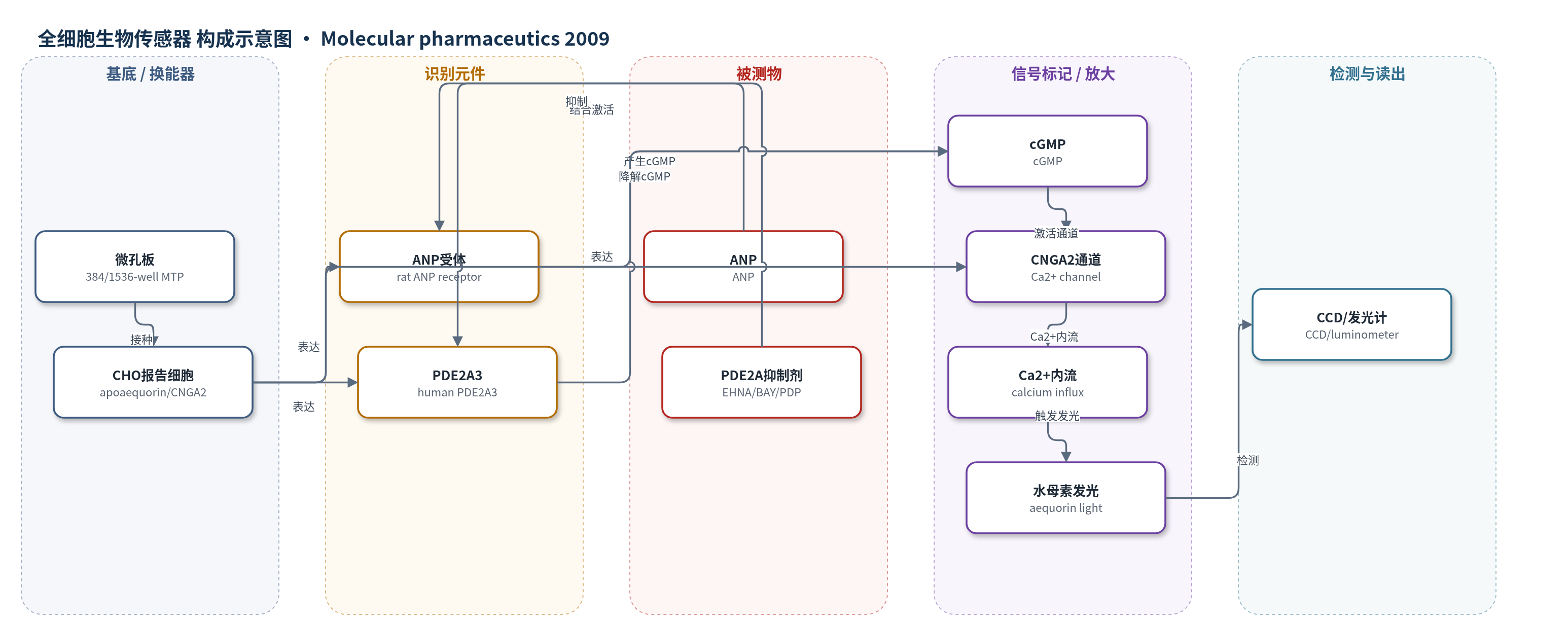
点击图片查看大图 · 依据论文自动绘制
传感器类型
全细胞生物传感器
检测对象
PDE2A 抑制剂(PDE2A inhibitors,如 EHNA、BAY 60-7550、PDP、IBMX、trequinsin、dipyridamole);样品基质为无钙 Tyrode 刺激液(含 0.1% BSA)中的化合物溶液,用于 CHO 报告细胞检测。
检测原理
ANP 结合细胞表面 ANP 受体并激活鸟苷酸环化酶,使胞内 cGMP 升高;稳定表达的人源 PDE2A3 随即水解 cGMP,使基础 cGMP 维持在低水平。当 PDE2A 抑制剂(如 EHNA、BAY 60-7550、PDP)进入细胞并结合 PDE2A3 时,cGMP 水解被抑制,cGMP 浓度随抑制剂浓度升高而累积。累积的 cGMP 激活 CNGA2 阳离子通道,引起 Ca2+ 内流;Ca2+ 与 apoaequorin/coelenterazine 复合物结合,触发化学发光。发光强度与 cGMP 水平及钙内流量正相关,因此抑制剂浓度越高,ANP 刺激下的发光越强,浓度-响应曲线左移。无 ANP 时,因缺乏 cGMP 生成,抑制剂不增加基础发光。实时模式在含 2 mM Ca2+ 的 Tyrode 中进行,可连续监测 cGMP 生成动力学。
检测灵敏度
未报告 LOD、线性范围、灵敏度斜率或相关系数。
效应效果
该细胞系选择性监测 PDE2A 抑制:亲代 ANP 细胞中 EHNA、BAY 60-7550、PDP 不改变 ANP 发光;PDE2A 细胞中增强信号并左移。10 nM ANP 下 pEC50 为 5.76±0.14、8.24±0.02、7.40±0.03;100 nM 下为 5.78±0.12、8.22±0.05、7.40±0.06。IBMX、trequinsin、dipyridamole 在 10 nM ANP 下 pEC50 为 4.13±0.05、5.32±0.12、5.19±0.02。其他 PDE 抑制剂无信号。PDP 细胞活性较生化 IC50 0.6 nM 低约 60 倍,Papp A→B 2.69×10^-7 cm/s,外排比 64.4;BAY 为 1.30×10^-5 cm/s。1 µM BAY 使动力学信号增强约 130 倍,可转移至 1536 孔 uHTS。
传感器的构成
- 检测载体:不透明 384 孔微孔板(opaque 384-well MTP)或 1536 孔微孔板(1536-well MTP),用于细胞培养与发光检测。
- 换能细胞基质:重组 CHO 细胞系(recombinant CHO cell line),稳定表达 apoaequorin 与 CNGA2 通道,作为胞内 cGMP 传感与钙依赖发光换能单元。
- 识别/刺激元件:大鼠 ANP 受体(rat ANP receptor),识别 ANP 并激活鸟苷酸环化酶产生 cGMP。
- 靶酶/识别元件:人源 PDE2A3(human PDE2A3),降解 cGMP,作为 PDE2A 抑制剂的结合靶点。
- 信号通道:CNGA2 通道(CNGA2 channel),被 cGMP 激活后允许 Ca2+ 内流。
- 发光标记物:apoaequorin(水母素蛋白)与 coelenterazine(腔肠素),在 Ca2+ 存在下发生化学发光。
- 刺激/缓冲介质:无钙 Tyrode 溶液(Ca2+-free Tyrode,含 130 mM NaCl、5 mM KCl、20 mM HEPES、1 mM MgCl2、4.8 mM NaHCO3、pH 7.4)及 0.1% BSA;实时模式含 2 mM Ca2+。
- 读出装置:CCD 相机(CCD camera,Hamamatsu)或常规发光计(luminometer),用于实时或积分发光信号读出。
中文摘要
本文报道了一种磷酸二酯酶2A(PDE2A)报告细胞系的构建与药理学表征。人源 PDE2A 被稳定转染至表达心房利钠肽(ANP)受体和环核苷酸门控阳离子通道 CNGA2 的亲代细胞系中,CNGA2 作为胞内 cGMP 生物传感器。该细胞系可通过钙离子经 CNG 通道内流刺激水母素发光,实时监测 cGMP 水平。研究显示,该检测能以高灵敏度特异性监测 PDE2A 抑制。无 ANP 刺激时,选择性抑制剂 EHNA、BAY 60-7550 和 PDP 不增加基础发光;与 ANP 联用时则增强发光并使 ANP 浓度-响应曲线左移。非选择性抑制剂 IBMX、trequinsin 和 dipyridamole 也呈类似结果。最强 PDE2A 抑制剂 PDP 的细胞活性低于生化预期,转运研究显示其通透性低且为外排转运体底物。其他 PDE 抑制剂未刺激发光。结果表明该 PDE2A 报告检测可高效、高通量地鉴定和表征 PDE2A 抑制剂。
英文摘要
We report here the generation and pharmacological characterization of a phosphodiesterase 2A (PDE2A) reporter cell line. Human PDE2A was stably transfected in a parental cell line expressing the atrial natriuretic peptide (ANP) receptor and the cyclic nucleotide-gated (CNG) cation channel CNGA2, acting as the biosensor for intracellular cGMP. In this reporter cell line, cGMP levels can be monitored in real-time via aequorin luminescence stimulated by calcium influx through the CNG channel. By using different PDE inhibitors, we could show that our PDE2A reporter assay specifically monitors PDE2A inhibition with high sensitivity. In the absence of ANP stimulation, the PDE2A selective inhibitors EHNA, BAY 60-7550 and PDP did not increase basal luminescence levels in this experimental setting. However, in combination with ANP, these inhibitors stimulated luminescence signals and induced leftward shifts of ANP concentration-response curves. Similar results were obtained when using IBMX, trequinsin and dipyridamole, which inhibit PDE2A nonselectively with lower potency. PDP, the most potent PDE2A inhibitor known to date, was found to exhibit much lower cellular activity as anticipated from its biochemical PDE2A inhibitory activity. By cellular uptake and transport studies we could show that PDP's cell permeability is low and that the compound is a substrate for an efflux transporter. Other PDE inhibitors including vinpocetine, milrinone, rolipram, sildenafil, zaprinast, BRL 50481 and BAY 73-6691 did not stimulate luminescence signals on our PDE2A reporter cell line. The results imply that this novel PDE2A reporter assay provides an efficient, high throughput means for the identification and characterization of PDE2A inhibitors.