组成图示
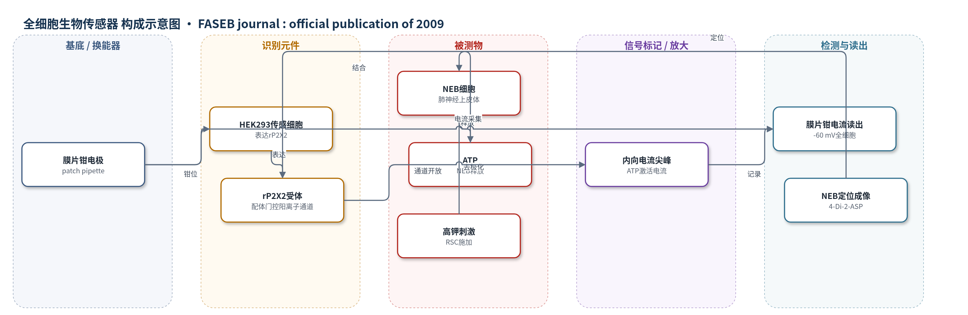
点击图片查看大图 · 依据论文自动绘制
传感器类型
全细胞生物传感器
检测对象
ATP(三磷酸腺苷),样品基质为离体小鼠肺切片(ex vivo mouse lung slices)中NEB微环境
检测原理
高钾溶液经快速换液系统施加于离体肺切片中的NEB,使NEB细胞去极化并触发钙依赖的量子化ATP释放。释放的ATP在NEB微环境中短距离扩散,到达邻近的HEK293传感细胞。该细胞膜上稳定表达大鼠P2X2受体(rP2X2),ATP结合后使配体门控阳离子通道开放,Na+和Ca2+内流。在全细胞膜片钳钳位电位-60 mV下,离子内流被记录为短暂内向电流尖峰。电流尖峰的出现、幅度和频率反映局部ATP释放事件与浓度变化。该方法不依赖酶促或核酸放大,其选择性主要来自rP2X2对ATP的识别以及传感细胞与NEB之间数微米的空间接近;若传感细胞移离NEB约10 μm,则无法检测到ATP释放。
检测灵敏度
原文未报告LOD、线性范围、灵敏度斜率或相关系数。
效应效果
该reporter-patching ATP生物传感器表现出高空间选择性和时间分辨率。只有当表达rP2X2的HEK293细胞被置于NEB下游气道上皮表面附近时,高钾刺激才能诱导向内电流尖峰;将传感细胞移离NEB约10 μm后,高钾刺激不再产生电流,说明检测局限于NEB微环境。ATP阳性对照(10 μM,2 s)可稳定诱发内向电流,溶液交换到检测的时间滞后约1 s。生物学验证中,NEB去极化后Clara样细胞二次激活比例由94%降至57%(apyrase/腺苷脱氨酶)或7%(suramin),支持ATP/P2受体介导的旁分泌信号。作者认为该方法为直接检测离体组织局部ATP释放提供了灵敏、选择性工具。
传感器的构成
- 换能器电极:全细胞膜片钳电极(patch pipette),用于钳位传感细胞并采集内向电流
- 传感细胞:HEK293细胞,稳定表达大鼠P2X2嘌呤能受体(rP2X2),作为ATP识别与离子通道换能元件
- 识别元件:rP2X2受体,配体门控阳离子通道,结合ATP后开放并产生内向电流
- 样品基质:离体小鼠肺切片(ex vivo mouse lung slices),NEB位于气道上皮微环境
- 释放触发:高钾溶液(high K+)经多管快速换液系统(RSC)施加,使NEB去极化并释放ATP
- 定位标记:4-Di-2-ASP荧光染料,标记NEB以便在切片中定位传感细胞
- 信号读出:膜片钳内向电流尖峰(inward current spikes),反映ATP到达传感细胞并激活rP2X2
中文摘要
肺神经上皮体(NEB)是富含神经支配的复杂气道感受器,参与呼吸调节,并与周围Clara样细胞共同具有干细胞潜能。本研究采用共聚焦活细胞成像和新型电生理技术,在离体肺切片模型中解析NEB微环境中的嘌呤能信号通路。奎那克林组织化学显示NEB细胞富含囊泡ATP。作者将reporter-patching方法改造为高灵敏、高选择性ATP生物传感器,直接检测离体NEB去极化后ATP的量子化释放。增强胞外ATP酶解或阻断P2受体证实ATP在NEB细胞与Clara样细胞旁分泌相互作用中的核心作用。钙成像、药理学和免疫组织化学表明,ATP与功能性P2Y2受体结合介导Clara样细胞激活。因此,NEB细胞通过释放ATP与邻近细胞通讯,并可能影响气道功能、损伤后上皮再生及小细胞肺癌发生。
英文摘要
Pulmonary neuroepithelial bodies (NEBs) are densely innervated groups of complex sensory airway receptors involved in the regulation of breathing. Together with their surrounding Clara-like cells, they exhibit stem cell potential through their capacity to regenerate depopulated areas of the epithelium following lung injury. We have employed confocal live cell imaging microscopy and novel electrophysiological techniques in a new ex vivo lung slice model to unravel potential purinergic signaling pathways within the NEB microenvironment. Quinacrine histochemistry indicated high amounts of vesicular ATP in NEB cells. Using a "reporter-patching" method adapted to create a uniquely sensitive and selective biosensor for the direct detection of ATP release from NEBs ex vivo, we demonstrated quantal ATP release from NEBs following their depolarization. Enhancing enzymatic extracellular ATP hydrolysis or inhibiting P2 receptors confirmed the central role of ATP in paracrine interactions between NEB cells and Clara-like cells. Combined calcium imaging, pharmacology, and immunohistochemistry showed that ligand-binding to functional P2Y(2) receptors underpins the activation of Clara-like cells. Hence, NEB cells communicate with their cellular neighbors in the NEB microenvironment by releasing ATP, which rapidly evokes purinergic activation of surrounding Clara-like cells. Besides ATP acting on the P2X(3) receptor expressing vagal sensory nerve terminals between NEB cells, local paracrine purinergic signaling within this potential stem cell niche may be important to both normal airway function, airway epithelial regeneration after injury, and/or the pathogenesis of small cell lung carcinomas.