组成图示
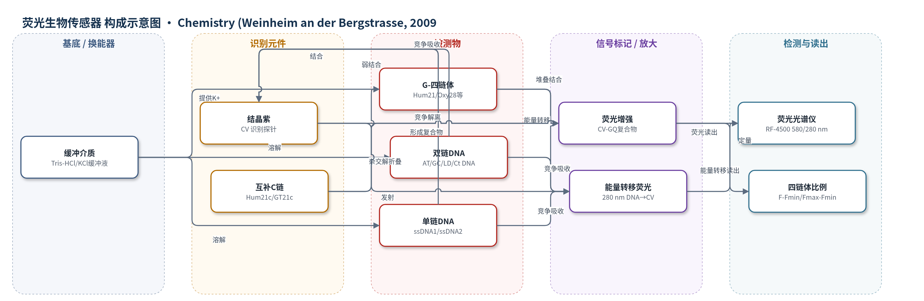
点击图片查看大图 · 依据论文自动绘制
传感器类型
荧光生物传感器
检测对象
G-四链体(G-quadruplex)、双链DNA(duplex DNA)、单链DNA(ssDNA);样品基质为 Tris-HCl/KCl 缓冲液中的寡核苷酸溶液
检测原理
CV 与 G-四链体结合时,其三个苯环堆叠到外侧 G-四分体,二甲氨基与磷酸骨架及环区发生静电/氢键作用,染料构象刚性增加,荧光显著增强;双链或单链 DNA 仅引起较弱增强或竞争吸收导致荧光下降。在 280 nm 激发下,DNA 吸收能量并转移至结合态 CV,产生能量转移荧光;G-四链体使该信号增强,过量 DNA 竞争吸收则使信号下降。固定 CV 浓度滴定 DNA 时,G-四链体曲线出现拐点,可确定 2:1 化学计量和结合常数。加入 C 富互补链后,G-四链体解折叠为双链,CV 荧光下降,按 (F-Fmin)/(Fmax-Fmin) 计算四链体比例。
检测灵敏度
结合常数 Ka: (0.036±0.008) μM−2 (Hum21)、(0.019±0.003) μM−2 (Oxy28)、(0.0033±0.0008) μM−2 (Hum12)、(0.0066±0.0003) μM−2 (Oxy12);结合化学计量比: 2:1
效应效果
CV 对分子内 G-四链体(Hum21、Oxy28)结合优先,对分子间 G-四链体(Hum12、Oxy12)中等,对短双链(AT、LD、GC)弱或无结合;但竞争透析显示 CV 也可结合 ssDNA 和长双链 Ct DNA,水平与分子间四链体相当。能量转移荧光比荧光更能清晰区分结构并确定 2:1 化学计量。与 G-四链体分子信标法相比,CV 法测得 Hum21 在 1:1 互补链时约 20% 四链体,GT21 约 78%;信标法分别为约 16% 和 63%。CD 表明 CV 不显著改变四链体结构。作者认为该方法可用于区分 DNA 结构、比较四链体形成能力、测定互补序列下四链体比例及监测结构变化。
传感器的构成
- 检测缓冲层:25 mM Tris-HCl(pH 7.0)/100 mM KCl,提供 K+ 稳定 G-四链体并维持荧光反应
- 识别/信号探针:结晶紫(CV),三苯甲烷荧光染料,结合 G-四链体后荧光增强,作为识别与信号元件
- 辅助对照探针:甲基绿(MG),三苯甲烷荧光染料,用于能量转移荧光比较
- 竞争识别元件:C 富互补链(Hum21c、GT21c),与 G 链杂交竞争四链体形成,用于定量四链体比例
- 读出装置:Hitachi RF-4500 荧光光谱仪,检测 580 nm 激发荧光或 280 nm 激发能量转移荧光
中文摘要
富含鸟嘌呤的核酸序列常存在于端粒、基因启动子等生物重要区域,且多与互补链共存,因此发展灵敏的生物传感器以区分 G-四链体与双链结构并测定四链体相对双链的竞争能力备受关注。本研究采用荧光光谱和能量转移荧光光谱,考察了两种三苯甲烷染料甲基绿(MG)和结晶紫(CV)与 G-四链体、双链 DNA 或单链 DNA 的相互作用。结果表明,利用 CV 的荧光光谱或 CV 与 MG 的能量转移荧光光谱,可良好区分 G-四链体与双链或单链 DNA。通过 CV 与 G-四链体的能量转移荧光滴定,可确定 CV 与 G-四链体的结合化学计量比;通过 G-四链体–CV 复合物与 C 富互补链的荧光滴定,可测量存在互补序列时 G 富寡核苷酸参与 G-四链体结构的分数。该研究为区分四链体与双链或单链 DNA,以及测定互补 C 富序列存在下的 G-四链体比例提供了简便方法。
英文摘要
G-rich nucleic acid sequences with the potential to form G-quadruplex structures are common in biologically important regions. Most of these sequences are present with their complementary strands, so the development of a sensitive biosensor to distinguish G-quadruplex and duplex structures and to determine the competitive ability of quadruplex to duplex structures has received a great deal of attention. In this work, the interactions between two triphenylmethane dyes (malachite green (MG) and crystal violet (CV)) and G-quadruplex, duplex, or single-stranded DNAs were studied by fluorescence spectroscopy and energy-transfer fluorescence spectroscopy. Good discrimination between quadruplexes and duplex or single-stranded DNAs can be achieved by using the fluorescence spectrum of CV or the energy-transfer fluorescence spectra of CV and MG. In addition, by using energy-transfer fluorescence titrations of CV with G-quadruplexes, the binding-stoichiometry ratios of CV to G-quadruplexes can be determined. By using the fluorescence titrations of G-quadruplex-CV complexes with C-rich complementary strands, the fraction of G-rich oligonucleotide that engages in G-quadruplex structures in the presence of the complementary sequence can be measured. This study may provide a simple method for discrimination between quadruplexes and duplex or single-stranded DNAs and for measuring G-quadruplex percentages in the presence of the complementary C-rich sequences.