组成图示
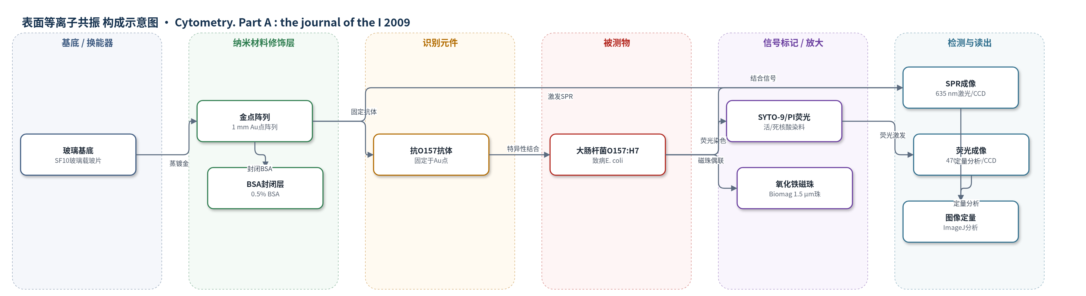
点击图片查看大图 · 依据论文自动绘制
传感器类型
表面等离子共振(SPR)生物传感器
检测对象
致病大肠杆菌 O157:H7(Escherichia coli O157:H7),样品基质为经磁珠预浓缩的细菌悬液/水样(PBS/水稀释)
检测原理
该装置先以偶联抗O157:H7抗体的氧化铁磁珠从样品中捕获并磁浓缩目标菌,提高微流控进样的采样统计。金点阵列表面固定抗O157:H7抗体,目标菌与抗体特异性结合后,金-介质界面附近的质量/介电性质改变,使Kretschmann构型下p偏振光激发的表面等离子体共振条件发生偏移,反射光强度或共振角变化被CCD成像记录。同时,细菌经SYTO-9和PI染色,顶部倒置荧光成像分别给出总菌数和膜损伤死菌信息。结合菌数越多,SPR反射变化区域和荧光覆盖面积越大;ImageJ按金点面积上细菌覆盖比例定量,从而同时获得病原存在、数量与相对感染性。
检测灵敏度
未报告LOD、线性范围、灵敏度斜率或R^2。
效应效果
磁珠-细菌复合物经磁场回收后,1:10至1:100稀释样的回收率为91%、93%、92.4%和95%,均大于90%。在4×4金点阵列中,仅抗O157:H7抗体点接触O157:H7时出现显著结合,平均结合1653个菌;兔非免疫血清、BSA点及DH5-a对照点平均结合少于300个菌,表明非特异结合低。捕获菌经SYTO-9/PI成像显示活力为97.7%。系统可在高低菌密度下对单个致病大肠杆菌进行SPR和荧光成像,并可用4×4阵列最多实现16个功能化点、约14种病原加对照的多路筛查。作者认为该便携混合成像设备可现场、无标记、实时检测食源性病原,避免传统培养数天的耗时。
传感器的构成
- 基底/换能器:玻璃载玻片(glass slide/SF10 glass)与1 mm直径金点阵列(gold spots, 4×4 array),提供SPR换能表面并固定识别分子。
- 识别元件:抗大肠杆菌O157:H7特异性抗体(anti-E. coli O157:H7 antibody, 100 μg/mL),点样固定于金点,特异性捕获目标菌。
- 负对照/封闭:兔非免疫血清(rabbit preimmune serum)和0.5%牛血清白蛋白(BSA Fraction V),分别作阴性对照并封闭金表面及抗体非特异位点。
- 磁浓缩捕获元件:超顺磁性羧基功能化氧化铁微珠(superparamagnetic carboxyl-functionalized Biomag 1.5 μm iron oxide beads)偶联抗O157:H7抗体,用于从大体积样品中磁捕获和富集细菌。
- 荧光标记物:SYTO-9核酸染料与碘化丙啶(PI),标记细菌用于总菌数与死菌/活菌状态的倒置荧光成像。
- 微流控层:聚二甲基硅氧烷(PDMS)微流控流室,封装金点阵列并输送磁浓缩样品。
- 光学读出组件:Kretschmann构型棱镜(SFL11/BK7 prism)、635 nm激光二极管、偏振器、CCD相机及470 nm LED/滤光片,用于SPR与epi-fluorescence成像。
中文摘要
现有食品病原筛查方法依赖昂贵抗体或PCR试剂,或需耗时培养。为满足便携、实时、多路复用病原检测需求,作者设计了一种混合微流控生物芯片,将表面等离子共振(SPR)成像与荧光成像结合,用于单细胞病原的多路检测。该芯片由金点阵列构成,每个金点固定针对特定病原的捕获生物分子,并由聚二甲基硅氧烷(PDMS)微流控流室封装,用于输送经磁浓缩的待测样品。样品在芯片底部接受SPR成像,在顶部接受倒置荧光成像。原型系统成功通过SPR和荧光成像观察抗体捕获的大肠杆菌O157:H7。利用氧化铁在350 nm处的吸光度测定磁珠捕获效率,并用NIH ImageJ软件按金点面积上细菌覆盖比例定量结合量。该混合成像方法可同时估计相对感染性,为食源性病原检测提供了可行原型。
英文摘要
Current methods to screen for bacterial contamination involve using costly reagents such as antibodies or PCR reagents or time-costly growth in cultures. There is need for portable, real-time, multiplex pathogen detection technology that can predict the safety of food. Surface plasmon resonance (SPR) imaging is a sensitive, label-free method that can detect the binding of an analyte to a surface by the changes in refractive index that occur upon binding. We have designed a hybrid microfluidic biochip to perform multiplexed detection of single-celled pathogens using a combination of SPR and fluorescence imaging. The device consists of an array of gold spots, each functionalized with a capture biomolecule targeting a specific pathogen. This biosensor array is enclosed by a polydimethylsiloxane microfluidic flow chamber that delivers a magnetically concentrated sample to be tested. The sample is imaged by SPR on the bottom of the biochip and epi-fluorescence on the top. The prototype instrument was successfully able to image antibody-captured E. coli O157:H7 bacteria by SPR and fluorescence imaging. The efficiency of capture of these bacteria by the magnetic particles was determined using spectrophotometric ferric oxide absorbance measurements. The binding of the E. coli to each spot was quantified by measuring the percent of the gold spot area upon which the bacteria was bound and analyzed using NIH ImageJ software. This hybrid imaging approach of pathogenic E. coli detection coupled with an estimate of relative infectivity is shown to be a working example of a testing device for potential foodborne pathogens.