组成图示
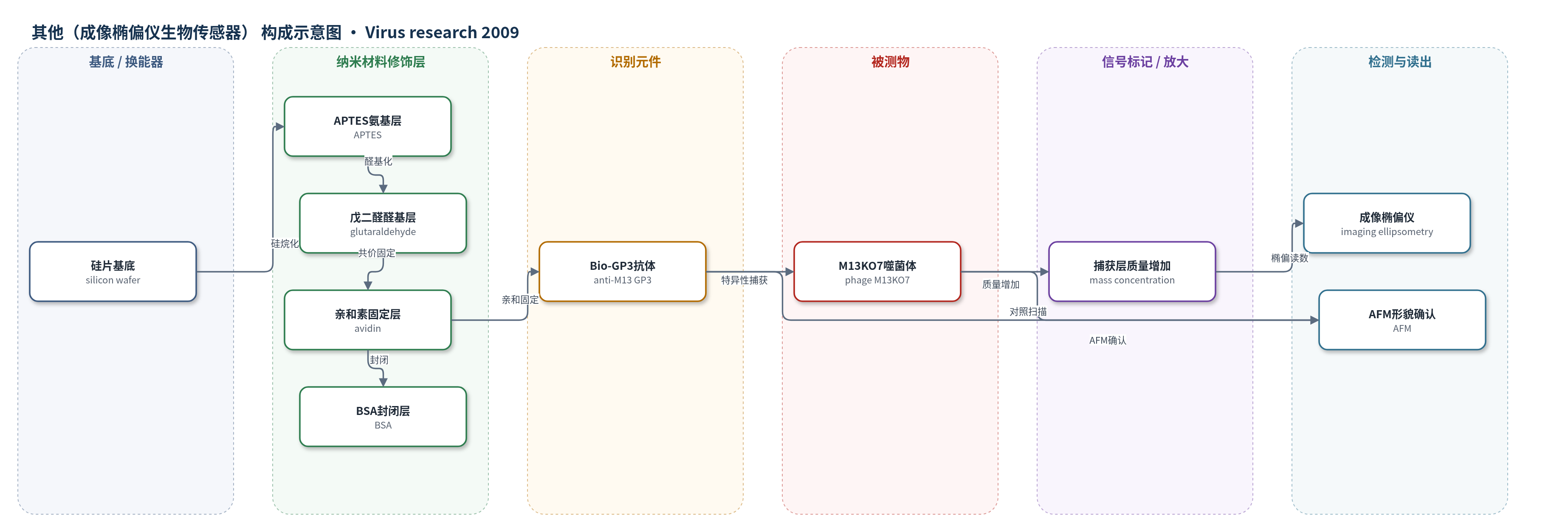
点击图片查看大图 · 依据论文自动绘制
传感器类型
其他(成像椭偏仪生物传感器)
检测对象
噬菌体M13KO7(phage M13KO7);样品基质为噬菌体培养液、纯化噬菌体样品及PBST稀释液,另以SARS病毒作阴性对照
检测原理
硅片经APTES和戊二醛修饰形成醛基,avidin通过席夫碱共价固定,BSA封闭后,Bio-GP3经avidin-biotin定向固定,Fab端朝外识别M13KO7的pIII小衣壳蛋白。当噬菌体溶液流过传感面时,M13KO7被抗体特异性捕获,表面质量浓度和等效膜厚增加。成像椭偏仪以偏振光探测薄膜光学特性,反射/透射光携带层厚信息;在0–30 nm范围内灰度值I与膜厚d满足I=kd^2,表面浓度≈K×d(K=0.12),因此灰度随捕获噬菌体量增加而升高。该过程无标记、无酶放大,AFM用于微观形貌确认。
检测灵敏度
灵敏度: 10^9 pfu/ml;分辨率: 0.7 × 10^9 pfu/ml;表1检测浓度: 0–2.5 × 10^11 pfu/ml,对应表面浓度: 0–2.1 μg/cm2
效应效果
BIE可区分不同批次噬菌体:第一批灰度变化约151%,第二批约2%,提示前者有活性、后者失活或无噬菌体。纯化M13KO7重复检测中,avidin浓度0.5、1、1.25 mg/ml产生不同灰度。SARS阴性对照无显著表面浓度增加,显示选择性。90 Hz超声分散聚集体,使检测区均匀。AFM确认:GP3约98.2±0.14 nm长、1.7±0.46 nm高、92.6±0.19 nm宽;捕获M13KO7长丝约1 μm、高4.42±0.173 nm、宽39.21±0.30 nm。作者认为BIE可无标记检测完整病毒并支持多路并行,但灵敏度10^9 pfu/ml低于AFM/QCM的10^8 pfu/ml。
传感器的构成
- 基底/换能器:硅片(silicon wafer),经H2O2:H2SO4清洗形成硅羟基,作为成像椭偏仪检测基底
- 硅烷化氨基层:3-氨基丙基三乙氧基硅烷(APTES)与硅羟基共价结合,形成表面氨基
- 醛基交联层:戊二醛(glutaraldehyde)与氨基反应生成醛基(-CHO),用于共价固定蛋白
- 亲和固定层:亲和素(avidin)通过席夫碱与醛基共价固定,提供生物素结合位点
- 封闭层:牛血清白蛋白(BSA)封闭非特异结合位点
- 识别元件:生物素标记抗M13KO7 GP3抗体(Bio-GP3)经avidin-biotin定向固定,Fab端朝外识别pIII小衣壳蛋白
- 微流控图案化:微流控系统(microfluidic system)实现蛋白阵列图案化与样品输送;超声分散噬菌体聚集体
- 信号标记/放大:无外源标记,M13KO7捕获后增加表面质量浓度/膜厚,成像椭偏仪灰度变化作为信号
- 读出头:成像椭偏仪(imaging ellipsometry)测量灰度/表面浓度;AFM用于形貌确认
中文摘要
病原体的快速检测与鉴定对减少疾病传播具有重要意义。本研究开发了一种基于成像椭偏仪(BIE)的无标记、多路生物传感器,用于检测噬菌体M13KO7。硅片表面经醛基修饰,并通过微流控系统使蛋白质以阵列形式在硅片表面均匀、同时图案化。亲和素(avidin)被固定在表面,利用亲和素与生物素的高亲和力实现生物素标记抗M13抗体(Bio-GP3)的定向固定,作为识别噬菌体M13KO7的配体。当含M13KO7的溶液流过表面时,噬菌体被配体特异性捕获,导致抗体-噬菌体结合层的表面质量浓度显著增加,成像椭偏仪可检测到该变化,灵敏度为10^9 pfu/ml。此外,原子力显微镜(AFM)进一步证实噬菌体M13KO7已被表面配体直接捕获。结果表明,BIE可用于M13KO7的直接检测,并在病毒检测领域具有应用潜力。
英文摘要
A rapid detection and identification of pathogens is important for minimizing transfer and spread of disease. A label-free and multiplex biosensor based on imaging ellipsometry (BIE) had been developed for the detection of phage M13KO7. The surface of silicon wafer is modified with aldehyde, and proteins can be patterned homogeneously and simultaneously on the surface of silicon wafer in an array format by a microfluidic system. Avidin is immobilized on the surface for biotin-anti-M13 immobilization by means of interaction between avidin and biotin, which will serve as ligand against phage M13KO7. Phages M13KO7 are specifically captured by the ligand when phage M13KO7 solution passes over the surface, resulting in a significant increase of mass surface concentration of the anti-M13 binding phage M13KO7 layer, which could be detected by imaging ellipsometry with a sensitivity of 10(9)pfu/ml. Moreover, atomic force microscopy is also used to confirm the fact that phage M13KO7 has been directly captured by ligands on the surface. It indicates that BIE is competent for direct detection of phage M13KO7 and has potential in the field of virus detection.