组成图示
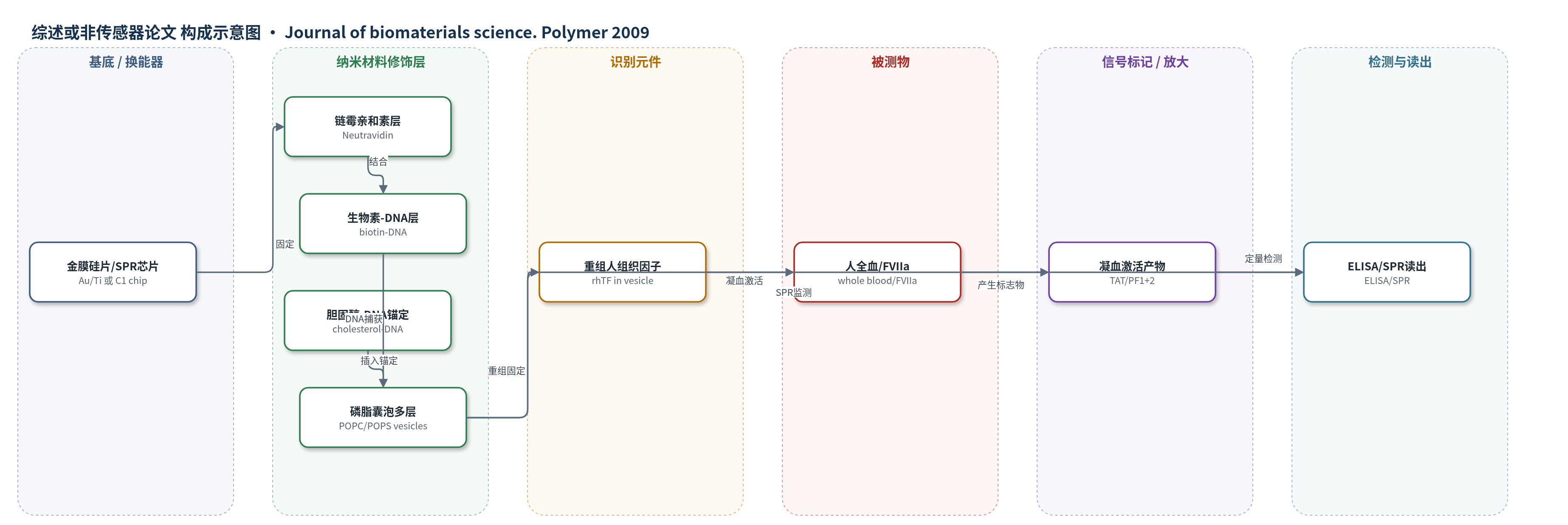
点击图片查看大图 · 依据论文自动绘制
传感器类型
综述或非传感器论文
检测对象
人全血凝血激活(blood coagulation activation,以 TAT、PF1+2、rhTF 为标志物);样品基质:人全血(枸橼酸抗凝、复钙)
检测原理
该表面并非以单一识别-换能方式检测分析物,而是将 rhTF 重组于磷脂囊泡膜中,模拟细胞膜外源性凝血途径。胆固醇-DNA 锚定使囊泡通过互补 DNA 捕获在金表面形成多层脂质膜。全血流经表面时,rhTF 与血浆中的凝血因子 VIIa 相互作用,启动外源性凝血级联,生成凝血酶;凝血酶与抗凝血酶形成 TAT 复合物,并裂解凝血酶原产生 PF1+2。ELISA 定量这些标志物,其水平随表面促凝活性升高而升高。加入直接凝血酶抑制剂 melagatran 后,凝血酶活性被抑制,TAT 与 PF1+2 呈浓度依赖性下降。SPR 用于监测囊泡层组装质量。
检测灵敏度
原文未报告 LOD、线性范围、灵敏度斜率或相关系数。
效应效果
SPR 显示 4 层 rhTF 囊泡约 3000 RU,POPC/POPS 囊泡约 6000 RU;rhTF 囊泡直径约 70 nm、多分散性约 0.5,挤出囊泡约 100 nm、多分散性约 0.1。全血流动腔中,rhTF 表面使 TAT 升高 8.5 倍,高于 POPC(1.1 倍)、POPC/POPS(3.4 倍)和主动脉(3.5 倍);PF1+2 在 rhTF 修饰 POPC/POPS 表面升高 1.8 倍,主动脉为 1.5 倍,POPC 下降。血浆 rhTF 未因表面释放升高,表明稳定。melagatran 对 TAT 和 PF1+2 的 IC50 分别为 1.4 µmol/l 和 5.3 µmol/l,比主动脉参考方法高约一个数量级。作者认为可用于抗凝药和生物材料凝血活性表征。
传感器的构成
- 基底:金膜硅片或 Biacore C1 SPR 芯片,提供表面修饰与 SPR 检测基底(Au/Ti;C1 chip)。
- 连接/封闭层:biotin-BSA(0.1 mg/ml)用于流动腔金表面,或 amine coupling 固定 neutravidin(20 µg/ml)用于 SPR C1 芯片,形成生物素-链霉亲和素连接。
- DNA 捕获层:biotin-DNA(2 µmol/l)固定于链霉亲和素表面,提供互补 DNA 捕获位点。
- 锚定层:cholesterol-DNA tethers(10 µmol/l 或 0.4–0.5 µmol/l)插入磷脂囊泡膜,使囊泡可被表面 DNA 捕获。
- 囊泡层:POPC/POPS 磷脂囊泡或 rhTF 重组囊泡,形成 3–4 层多层脂质膜结构。
- 识别/功能元件:recombinant human tissue factor(rhTF)重组于囊泡膜中,模拟细胞膜外源性凝血激活位点。
- 检测标志物:TAT complex、PF1+2、rhTF,经 ELISA 定量(非表面标记物)。
中文摘要
本文报道了一种将重组人组织因子(rhTF)重组于磷脂囊泡中的新型表面固定化方法。rhTF 囊泡通过胆固醇-DNA 锚定分子自发插入脂质膜,并被固定在互补 DNA 链上,形成多层囊泡结构。采用表面等离子共振(SPR)生物传感器技术表征该 rhTF 囊泡表面修饰的性质。作为应用,作者将其用作血液凝固激活表面,并在 Badimon 流动腔中暴露于人全血以检测其促凝活性。血液流经流动腔后,血浆中 rhTF 水平未升高,表明 rhTF 表面修饰稳定。暴露于该表面后,凝血酶-抗凝血酶(TAT)复合物和凝血酶原片段 1+2(PF1+2)水平升高;加入直接凝血酶抑制剂 melagatran 后,二者呈浓度依赖性下降,说明 rhTF 触发的凝血活性可被抗凝抑制。该新型致血栓表面结果令人鼓舞,可进一步发展为凝血相关研究工具,例如抗凝药物和生物材料表征。
英文摘要
We describe a novel method to immobilize recombinant human tissue factor (rhTF) reconstituted in phospholipid vesicles. The rhTF vesicles were immobilized in a multilayer vesicle structure using cholesterol-DNA tethers spontaneously inserted into the lipid membrane. The properties of the rhTF vesicle surface modification were characterized by surface plasmon resonance biosensor technology. As an application of this surface modification, we investigated its use as a blood coagulation activating surface. The coagulation activating capacity of the surface modification was tested by exposure to human whole blood in a flow chamber. No increase in rhTF levels in the blood was found after passage through the flow chamber, indicating that the rhTF surface modification was stable. Thrombin-antithrombin (TAT) and prothrombin fragment (PF) 1 + 2 levels increased after exposure to the surface, and decreased in a concentration-dependent way upon addition of melagatran (a direct thrombin inhibitor), i.e., coagulation activity triggered by rhTF could be suppressed by anticoagulation. The results with this new thrombogenic surface are promising, and will be further developed into a useful tool for coagulation related investigations, e.g., characterization of anticoagulants and biomaterials.