组成图示
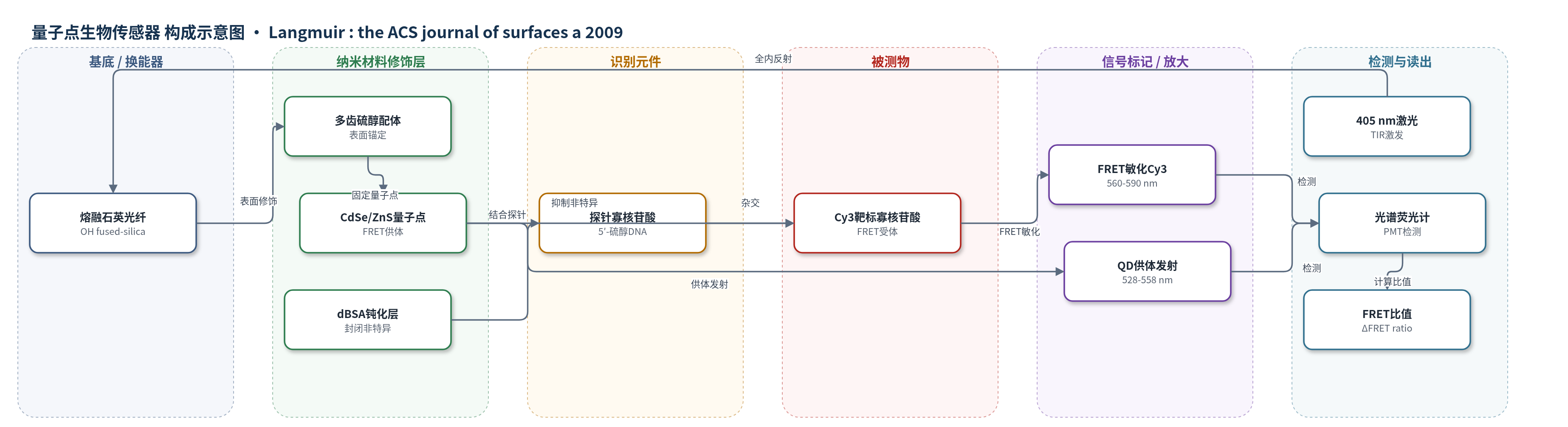
点击图片查看大图 · 依据论文自动绘制
传感器类型
量子点生物传感器
检测对象
互补单链DNA靶标寡核苷酸(Cy3-labeled target oligonucleotide)、三碱基错配DNA靶标(3-bp mismatched target);样品基质为PBS缓冲液中的寡核苷酸溶液
检测原理
光纤表面固定化 CdSe/ZnS 量子点(QD)作为 FRET 供体,探针寡核苷酸通过硫醇端连接于 QD 表面。当溶液中 Cy3 标记的互补靶标寡核苷酸与探针杂交时,Cy3 受体被拉近至 QD 附近(估算 Förster 距离约 3.5–4 nm),满足光谱重叠与距离条件。405 nm 激光经全内反射激发 QD,QD 发射能量非辐射转移至 Cy3,产生 FRET 敏化的 Cy3 荧光,同时 QD 发射被部分猝灭。以 560–590 nm Cy3 积分强度与 528–558 nm QD 积分强度之比作为 FRET 比值,扣除本底后 ΔFRET 比值随靶标浓度增加而增大。dBSA 钝化层封闭非特异性吸附位点,降低非互补序列假阳性。
检测灵敏度
LOD: 5 nM;动态范围: 接近2个数量级(almost 2 orders of magnitude);>250 nM时通常饱和
效应效果
dBSA 钝化后非互补 dA20 几乎无响应;0.1 µM 互补靶标 FRET 信号比非互补高 20–60 倍。未钝化时非互补信号可达靶标 50–100%。饱和靶标 FRET 效率约 30–50%,估算 Förster 距离 3.5–4 nm。25 °C 完全互补与三碱基错配信号比约 1.0:1.4,40 °C 为 1.0:0.3,50 °C 为 1.0:0.2,但互补信号较25°C下降67%。光纤可经 85:15 甲酰胺/PBS 再生并多次循环,但背景升高、信号下降,约 7 个循环后互补响应不再显著。作者认为性能与分子信标相当,动态范围优于此前溶液相约 1 个数量级。
传感器的构成
- 基底/换能器:熔融石英光纤(fused silica optical fiber),提供光导与全内反射激发平台
- 表面锚定层:多齿硫醇配体(multidentate thiol ligands),修饰光纤表面并固定量子点
- 量子点供体层:CdSe/ZnS 量子点(QDs,TOPO 包覆并经 MPA 配体交换),作为 FRET 供体
- 识别元件:5′-硫醇探针寡核苷酸(probe oligonucleotide,HS-C6H12-5′-ATT TTG TCT GAA ACC CTG T-3′),识别互补靶标
- 封闭/钝化层:变性牛血清白蛋白(dBSA,0.5 mg/mL PBS),阻断非特异性吸附
- 信号标记物:Cy3 标记靶标寡核苷酸(Cy3-labeled target oligonucleotide),作为 FRET 受体
中文摘要
本研究探索了以固定化量子点(QD)作为能量供体的荧光共振能量转移(FRET)方法,用于界面核酸杂交的换能检测。该工作源于 QD-FRET 在溶液相核酸杂交检测中的成功,并作为构建生物传感器的基础步骤,将选择性识别化学固定在固体表面。作者将 QD-探针寡核苷酸共轭物固定于光纤表面,随后引入 Cy3 标记的单链靶标寡核苷酸。杂交使 QD 供体与 Cy3 受体进入 FRET 有效距离,产生与靶标量成正比的荧光信号。结果表明,固相 QD-FRET 杂交检测可行;变性牛血清白蛋白(dBSA)钝化层可显著降低非特异性吸附,从而为可重复使用检测格式和错配判别提供可能。在该初步固相 QD-FRET 杂交检测中,检出限为 5 nM,动态范围接近两个数量级,并能利用三碱基错配序列区分完全互补靶标。尽管信号随循环逐渐下降,光纤仍可在多次杂交/解杂交循环中重复使用。研究还指出,通过优化 QD-探针寡核苷酸界面设计可进一步提升分析性能。
英文摘要
Fluorescence resonance energy transfer (FRET) using immobilized quantum dots (QDs) as energy donors was explored as a transduction method for the detection of nucleic acid hybridization at an interface. This research was motivated by the success of the QD-FRET-based transduction of nucleic acid hybridization in solution-phase assays. This new work represents a fundamental step toward the assembly of a biosensor, where immobilization of the selective chemistry on a surface is desired. After immobilizing QD-probe oligonucleotide conjugates on optical fibers, a demonstration of the retention of selectivity was achieved by the introduction of acceptor (Cy3)-labeled single-stranded target oligonucleotides. Hybridization generated the proximity required for FRET, and the resulting fluorescence spectra provided an analytical signal proportional to the amount of target. This research provides an important framework for the future development of nucleic acid biosensors based on QDs and FRET. The most important findings of this work are that (1) a QD-FRET solid-phase hybridization assay is viable and (2) a passivating layer of denatured bovine serum albumin alleviates nonspecific adsorption, ultimately resulting in (3) the potential for a reusable assay format and mismatch discrimination. In this, the first incarnation of a solid-phase QD-FRET hybridization assay, the limit of detection was found to be 5 nM, and the dynamic range was almost 2 orders of magnitude. Selective discrimination of the target was shown using a three-base-pairs mismatch from a fully complementary sequence. Despite a gradual loss of signal, reuse of the optical fibers over multiple cycles of hybridization and dehybridization was possible. Directions for further improvement of the analytical performance by optimizing the design of the QD-probe oligonucleotide interface are identified.