组成图示
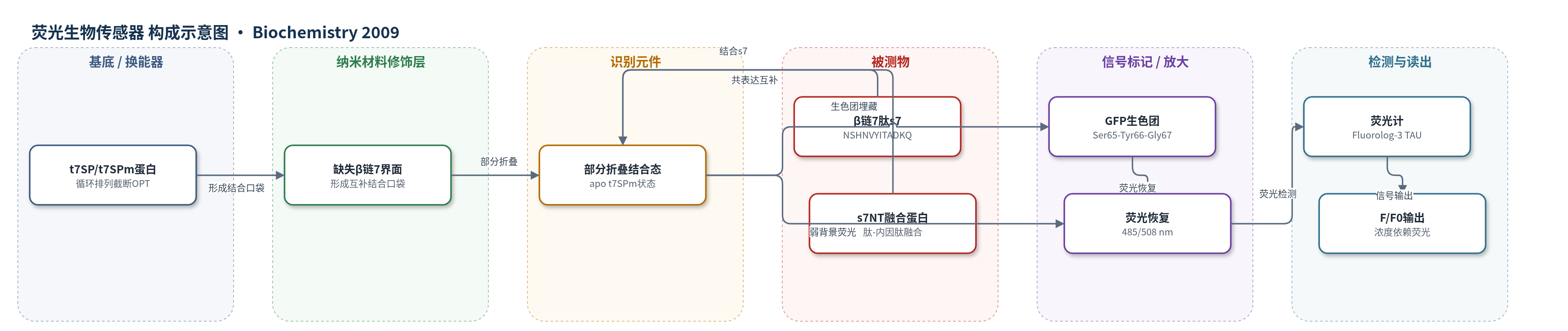
点击图片查看大图 · 依据论文自动绘制
传感器类型
荧光生物传感器
检测对象
β 链 7 肽(β-strand 7 peptide, s7, NSHNVYITADKQ)、肽-内因肽融合蛋白 s7NT(体内共表达来源);样品基质:体外 TN buffer(100 mM NaCl、50 mM Tris-HCl,pH 7.5)及大肠杆菌 BL21(DE3) 共表达细胞/细胞裂解物。
检测原理
该传感器基于蛋白片段互补与荧光自报告。循环排列并截断的 t7SP/t7SPm 缺少 β 链 7,其 β 桶部分开放,生色团部分暴露,因此仅显示弱荧光且生色团成熟缓慢。当游离 β 链 7 肽 s7 进入剩余 10 个 β 链形成的互补口袋并结合后,诱导 t7SPm 向天然态折叠,使 β 桶闭合、生色团被埋藏,从而恢复 485 nm 激发、508 nm 发射的荧光。荧光强度随 s7 浓度增加而增加,可用单位点结合等温线描述,Kd 约 0.5 µM。体系没有核酸或酶催化放大,信号增强主要来自肽结合驱动的折叠互补和生色团埋藏;预孵育肽可避免慢速新生色团形成,使荧光在约 10 min 内达到平台。体内 s7NT 通过内因肽剪接提供 s7 肽,共表达后同样恢复荧光。
检测灵敏度
Kd: approximately 5.51 × 10⁻⁷ M;corrected Kd: 5.31 × 10⁻⁷ M;abstract: ∼0.5 µM
效应效果
t7SP 与 s7NT 共表达使荧光较 t7SP 单独表达增加约 2 倍;t11SP/s11NT 可互补,t10SP/s10NT 无荧光,错配组合无额外荧光,显示序列特异性。体外 t7SPm 与 s7 结合后约 10 min 达到最终荧光的 90%,Kd 约 0.5 µM,比 src-SH3 的 3.0×10^-5 M 高约 100 倍。相对量子产率:t7SP 17.99±1.40%,t7SPm 39.50±0.27%,OPT 100±0.17%,c7SP 78.63±2.37%;互补 30 min 后接近 OPT。折叠为三态,5 min 内恢复 >50% 荧光;pKa 由 OPT 的 5.00 升至 8.08/8.22,酸性稳定性下降。高浓度 s7 下荧光衰减,可能源于 ROS/CALI。作者认为其可作自报告肽传感器。
传感器的构成
- 识别/换能蛋白:循环排列并截断的超级折叠 GFP OPT 变体 t7SP/t7SPm,缺少 β 链 7(residues 146-157),形成部分折叠结合口袋并自带荧光报告。
- 结合口袋:t7SP/t7SPm 剩余 10 个 β 链构成的互补界面,特异性结合缺失 β 链 7 肽 s7。
- 信号报告基团:GFP 生色团(由 Ser65-Tyr66-Gly67 环化氧化形成),肽结合后恢复 485/508 nm 荧光。
- 表达纯化辅助:6-His 标签与 Ni-NTA 亲和层析,用于制备纯化 t7SP/t7SPm。
- 反应介质:TN buffer(100 mM NaCl、50 mM Tris-HCl,pH 7.5),维持蛋白可溶与荧光测量。
中文摘要
绿色荧光蛋白(GFP)被用作一种新型“leave-one-out”生物传感器设计的概念验证:通过循环排列和截断使蛋白序列中部缺失一段,该蛋白可结合缺失肽并恢复功能。作者合成了三种 GFP 变体,分别缺失其 11 个 β 链中的第 7、10 或 11 链;其中两种变体在反式加入缺失肽序列后可恢复荧光。生化分析表明,缺失 β 链 7 的 t7SPm 处于部分去折叠状态,其 apo 形式与游离 β 链 7 肽结合,解离常数约 0.5 µM,并折叠为 GFP 天然态,从而恢复荧光。t7SPm 无论有无肽配体,折叠均为至少三态过程,速率与全长未排列 GFP 相当,提示循环排列和截断未改变折叠途径的限速步骤。该研究证明 GFP 的结构与功能可在链中部缺失一段时重建,且未结合形式处于部分去折叠状态。
英文摘要
Green fluorescent protein (GFP) has been used as a proof of concept for a novel "leave-one-out" biosensor design in which a protein that has a segment omitted from the middle of the sequence by circular permutation and truncation binds the missing peptide and reconstitutes its function. Three variants of GFP have been synthesized that are each missing one of the 11 beta-strands from its beta-barrel structure, and in two of the variants, adding the omitted peptide sequence in trans reconstitutes fluorescence. Detailed biochemical analysis indicates that GFP with beta-strand 7 "left out" (t7SPm) exists in a partially unfolded state. The apo form t7SPm binds the free beta-strand 7 peptide with a dissociation constant of approximately 0.5 microM and folds into the native state of GFP, resulting in fluorescence recovery. Folding of t7SPm, both with and without the peptide ligand, is at least a three-state process and has a rate comparable to that of the full-length and unpermuted GFP. The conserved kinetic properties strongly suggest that the rate-limiting steps in the folding pathway have not been altered by circular permutation and truncation in t7SPm. This study shows that structural and functional reconstitution of GFP can occur with a segment omitted from the middle of the chain, and that the unbound form is in a partially unfolded state.