组成图示
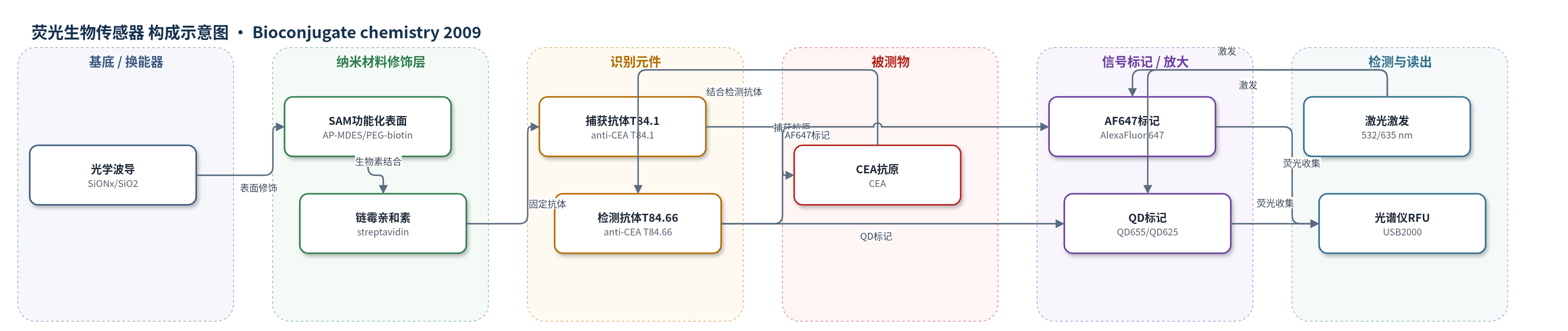
点击图片查看大图 · 依据论文自动绘制
传感器类型
荧光生物传感器
检测对象
癌胚抗原(carcinoembryonic antigen, CEA);样品基质:血清、乳头抽吸液(nipple aspirate fluid, NAF),文中另用50%牛血清评估非特异结合。
检测原理
该传感器采用平面光学波导的倏逝场进行空间过滤,仅激发距表面0–250 nm内的荧光分子,从而抑制血清等复杂样品中溶液相荧光背景。波导表面经SAM或PLB功能化,生物素—链霉亲和素相互作用固定捕获抗体T84.1;CEA与捕获抗体结合后,荧光标记检测抗体T84.66结合另一表位形成三明治复合物。激光激发AF647或QD,荧光发射经长通滤光片由光纤光谱仪收集为RFU。信号强度随CEA浓度增加而增加;AF647铰链区标记在重复激发下出现异常信号增强,可能源于多染料间去淬灭/能量转移,提高低浓度信背比;QD标记光稳定性高,适合长时间激发和多路检测。
检测灵敏度
LOD: <0.5 pM
效应效果
SAM表面检测100 pM CEA时S/B达483(3636/8 RFU),血清NSB 69 RFU、检测抗体NSB 193 RFU;未封闭PLB S/B仅44,封闭后74,SAM免封闭、耐洗涤且稳定>300天。重现性:25 pM CEA重复测量1046±119 RFU,患者样品跨波导外推78 pM、SD<1.5。AF647铰链区标记重复激发增强,100 pM初始2303 RFU(S/B 82),第4次5498 RFU(S/B 195);1 pM初始118 RFU,200 pM初始3033 RFU。QD标记100 pM时NH-QD655校正S/B约3、H-QD625约55,2 min激发无明显光漂白。作者认为可作即时诊断或研究工具,用于肿瘤标志物多路检测。
传感器的构成
- 基底/换能器:单模平面光学波导,抛光熔融石英基底+SiONx高折射率层+10 nm SiO2功能化层,表面刻衍射光栅耦合激光。
- 功能化表面:硅烷基自组装单分子层(SAMs),AP-MDES氨基硅烷化后接PEG-羧酸/甲氧基或保护氨基PEG,脱保护并接生物素;或DOPC+1% cap biotinyl磷脂双分子层(PLBs)。
- 固定层:链霉亲和素(streptavidin)与表面生物素结合,用于固定生物素化捕获抗体。
- 识别元件:生物素化捕获抗体T84.1结合CEA,检测抗体T84.66结合CEA另一表位,形成三明治免疫复合物。
- 信号标记物:检测抗体T84.66标记AF647(T84.66-NH-AF647或T84.66-H-AF647)或量子点QD655/QD625(T84.66-NH-QD655或T84.66-H-QD625)。
- 封闭/抗非特异:SAMs无需封闭;PLBs用PBS-2% BSA封闭;洗涤缓冲为PBS-0.5% BSA。
- 读出系统:532 nm激光激发QD、635 nm激光激发AF647,长通滤光片滤除激发光,USB2000光纤光谱仪收集荧光并输出RFU。
中文摘要
洛斯阿拉莫斯国家实验室传感器团队开发了一种基于平面光学波导的光学生物传感器,用于疾病相关生物标志物的敏感检测。此前该技术已用于乳腺癌患者血清和乳头抽吸液中癌胚抗原(CEA)的检测。本文报道了促进其向即时诊断系统或稳健研究工具转化的两项改进。首先,用硅烷基自组装单分子层(SAMs)替代磷脂双分子层(PLBs)进行波导表面功能化;SAMs 稳定、特异、耐洗涤,可减少非特异结合并提高信背比。其次,比较了四种荧光标记策略:检测抗体 T84.66 分别用 AlexaFluor 647(AF647)在赖氨酸氨基或铰链区标记,或用光稳定量子点(QDs)通过氨基或铰链区组氨酸标签标记。铰链区 AF647 标记在 635 nm 重复激发下出现异常信号增强,有利于低浓度抗原的信号分辨;量子点标记虽信号较低但抗光漂白能力显著优于有机染料。
英文摘要
The sensor team at the Los Alamos National Laboratory has developed a waveguide-based optical biosensor for the detection of biomarkers associated with disease. We have previously demonstrated the application of this technology to the sensitive detection of carcinoembryonic antigen in serum and nipple aspirate fluid from breast cancer patients. In this publication, we report improvements to this technology that will facilitate transition to a point-of-care diagnostic system and/or robust research tool. The first improvement involved replacing phospholipid bilayers used for waveguide functionalization with self-assembled monolayers. These thin films are stable, specific, and robust silane-based surfaces that reduce nonspecific binding and enhance the signal to background ratio. Second, we have explored four different fluorescent labeling paradigms to determine the optimal procedure for use in the assay. Labeling the detector antibody with an organic dye (AlexaFluor 647) in the hinge region allows for unusual signal enhancement with repeat excitation (at 635 nm) in our assay format, thereby facilitating a better signal resolution at lower concentrations of the antigen. We have also labeled the detector antibody with photostable quantum dots through either the amine groups of lysine (Fc, NH) or using a histidine tag in the hinge region of the antibody (Hinge, H). Both labeling strategies allow for acceptable signal resolution, but quantum dots show much greater resistance to photobleaching than organic dyes.