组成图示
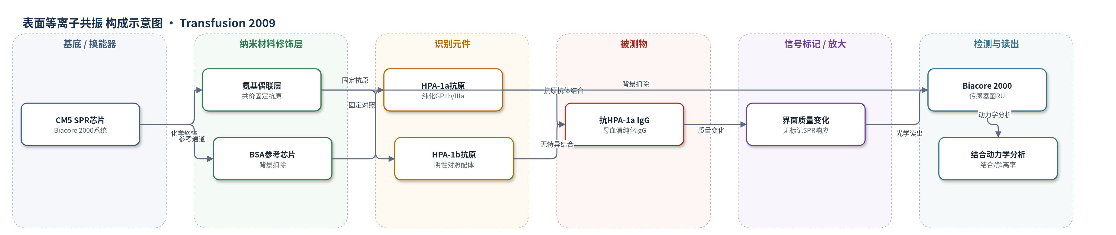
点击图片查看大图 · 依据论文自动绘制
传感器类型
表面等离子共振(SPR)生物传感器
检测对象
抗HPA-1a同种抗体(anti-HPA-1a alloantibody,IgG);样品基质:母体血清/血浆(经IgG纯化后)
检测原理
将免疫亲和纯化的HPA-1a或HPA-1b GPIIb/IIIa复合物通过氨基偶联固定于CM5 SPR芯片,形成抗原捕获表面。母体血清中纯化的IgG同种抗体随D-PBS流过芯片,抗HPA-1a IgG与固定HPA-1a结合,使界面结合质量改变,SPR仪实时记录共振单位(RU)传感器图。结合相反映抗体结合速率与浓度,解离相反映抗体解离速率;低亲和力抗体在结合相即逐渐解离,解离相后几乎完全脱落,高亲和力抗体则保持结合。由于SPR无需标记和洗涤,可避免MAIPA中反复洗涤导致低亲和力抗体丢失,从而检出MAIPA假阴性的低亲和力HPA-1a抗体。
检测灵敏度
未报告LOD、线性范围、灵敏度斜率或相关系数。
效应效果
SPR芯片对HPA-1a抗体特异:抗HPA-1a IgG仅结合HPA-1a芯片,不结合HPA-1b芯片;正常血清及含HPA-5b、HLA I类抗体的血清无响应。PTP抗体高响应约490 RU且几乎不解离;9例FNAIT抗体响应22.2–69.7 RU,解离后残留41%–79%。在2例MAIPA假阴性严重FNAIT病例中,SPR检出低亲和力HPA-1a抗体,结合相逐渐解离、解离相后完全脱落,说明MAIPA洗涤可去除此类抗体。作者认为SPR可辅助诊断临床相关低亲和力HPA-1a抗体,并可能评估母体抗体水平与新生儿血小板减少严重程度的关系。
传感器的构成
- 基底/换能器:CM5传感器芯片(Biacore 2000 SPR系统),提供SPR光学换能读出
- 修饰层:氨基偶联化学(amine-coupling),共价固定纯化抗原;乙醇胺封闭残余活性酯
- 识别元件:免疫亲和纯化的HPA-1a/HPA-1b GPIIb/IIIa复合物,作为固定配体捕获抗HPA-1a IgG
- 参考通道:BSA涂层芯片,用于扣除非特异背景信号
- 运行缓冲液:D-PBS(含0.005% P20),维持流动相并降低非特异结合
- 再生/清洗:25 mmol/L NaOH,解离结合抗体并再生芯片表面
中文摘要
胎儿和新生儿同种免疫性血小板减少症(FNAIT)多由母体抗人血小板抗原(HPA)-1a同种抗体引起。目前血清学诊断主要依赖单克隆抗体抗原捕获法(如MAIPA),但已有证据提示该方法可能漏检部分HPA-1a抗体。本研究采用表面等离子共振(SPR)技术,将免疫亲和纯化的糖蛋白IIb/IIIa(GPIIb/IIIa)HPA-1a/1b异构体固定于生物传感器芯片,实时分析不同FNAIT病例中HPA-1a同种抗体的结合动力学。结果显示,9例FNAIT血清中HPA-1a抗体呈中等相对响应(22.2–69.7共振单位,RU)且解离较慢,解离后芯片上仍残留41%–79%抗体;而输血后紫癜患者抗体在结合相末呈高响应(约490 RU)且几乎不解离。重要的是,SPR在2例MAIPA假阴性的严重FNAIT病例中检出HPA-1a抗体,其表现为结合相中逐渐解离、解离相后完全脱落,提示低亲和力抗体易被MAIPA的充分洗涤去除。结果表明,SPR有助于诊断临床相关的低亲和力HPA-1a抗体。
英文摘要
BACKGROUND: Fetal and neonatal alloimmune thrombocytopenia (FNAIT) is mostly caused by maternal alloantibodies directed against the human platelet alloantigen (HPA)-1a. Currently, the serologic diagnosis of FNAIT is based on the characterization of the HPA alloantibodies in monoclonal antibody-based antigen-capture assays (e.g., MAIPA assay). Accumulated current evidence indicated that such assays may overlook some HPA-1a antibodies.
STUDY DESIGN AND METHODS: This study employed surface plasmon resonance (SPR) technology using immunoaffinity-purified glycoprotein IIb/IIIa isoforms immobilized on biosensor chips to study the binding kinetics of HPA-1a alloantibodies from different FNAIT cases in real time.
RESULTS: Analysis of HPA-1a alloantibodies from FNAIT cases (n = 9) in SPR showed a moderate relative response (22.2-69.7 resonance units [RU]) and slow antibody dissociation. After the dissociation phase, varying amounts of bound antibodies (41%-79%) remained on the chip. In contrast in HPA-1a alloantibodies from a patient suffering from posttransfusion purpura, a high relative response (approximately 490 RU) was observed at the end of the association phase and no dissociation of antibody binding was detectable. Of particular relevance, by the use of this SPR technique, HPA-1a alloantibodies were detected in two severe FNAIT cases that had determined as false negative by MAIPA assay. In SPR, these HPA-1a alloantibodies showed low-avidity nature characterized by gradual dissociation of antibody during the association phase and complete detachment of antibody binding after the dissociation phase. This high "off-rate" character of low-avidity HPA-1a alloantibodies indicates that such antibody binding is easily detachable by the extensive washing procedure of the MAIPA.
CONCLUSIONS: Our results demonstrated that the SPR method can facilitate the diagnosis of clinically relevant low-avidity HPA-1a antibodies.