组成图示
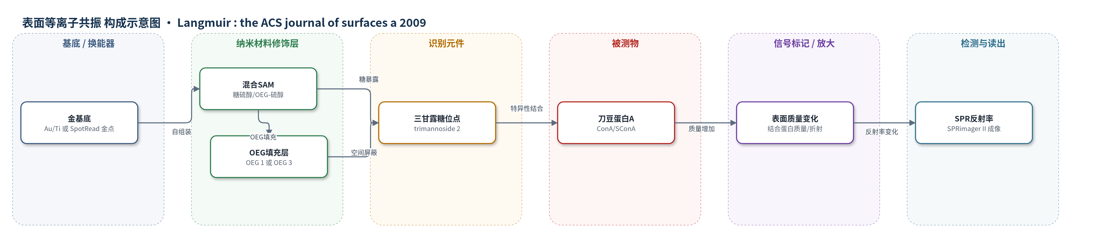
点击图片查看大图 · 依据论文自动绘制
传感器类型
表面等离子共振(SPR)生物传感器
检测对象
刀豆蛋白A(Concanavalin A, ConA)、琥珀酰刀豆蛋白A(succinyl-ConA, SConA);样品基质:PBS 缓冲液
检测原理
金表面通过 Au-S 键自组装糖硫醇与 OEG-硫醇混合 SAM,三甘露糖(trimannoside 2)暴露于溶液侧作为识别元件,OEG 链填充空隙并降低非特异吸附。当 PBS 中的刀豆蛋白 A(ConA)流过表面时,其多个甘露糖结合位点与表面三甘露糖特异性结合,使界面质量/折射率增加。SPR 换能机制为:结合质量改变金表面表面等离子体共振条件,SPRimager II 以反射率变化(normalized % reflectivity)实时无标记读出。信号随 ConA 浓度和表面糖密度增加而增强,但长 OEG 在糖浓度低于 60% 时因空间屏蔽使结合几乎消失;短 OEG 在 0–40% 糖浓度时结合显著上升,高糖密度后因拥挤略降。XPS C1s 中 O-C-O 缩醛峰用于定量糖覆盖,与 SPR 结合关联。
检测灵敏度
原文未报告 LOD、线性范围、灵敏度斜率或相关系数。报告数值:ConA 稀释至 12.5 nM (1.3 µg/mL);KADS: 3.1 ± 1.4 × 10^6 M^-1;KADS: 1.9 ± 1.1 × 10^6 M^-1;KADS: 1.3 ± 0.6 × 10^6 M^-1。
效应效果
SPR 成像显示 ConA 结合高度依赖表面糖密度和 OEG 链长。长 OEG 混合 SAM 中,糖浓度低于 60% 时 ConA 结合几乎不存在,100% 三甘露糖时结合最大;短 OEG 混合 SAM 中,0–40% 糖浓度时结合显著上升,40–60% 后出现平台并略降。SConA 与 ConA 的结合比约为 0.5,符合两者质量差异。RNase A 为阴性对照,RNase B 为阳性对照,OEG 背景扣除用于消除非特异信号。表面经 8 M 尿素再生 10 轮后仍保持 >95% 重现性。XPS C1s 缩醛 O-C-O 峰定量糖覆盖,比 O/C 比更准确,避免放射性标记。KADS 从 3.1 ± 1.4 × 10^6 M^-1 降至 1.3 ± 0.6 × 10^6 M^-1。作者认为 XPS 与 SPR 可用于糖基生物传感器/微阵列的表征与优化。
传感器的构成
- 基底/换能器:金膜(Au,Ti/Au 或 SpotRead 金点),提供 SPR 换能表面
- 识别元件:硫醇功能化三甘露糖(trimannoside 2, ManR1,2ManR1,2ManR-OEG-SH),经 Au-S 键固定并暴露甘露糖结合位点
- 混合填充层:长链 OEG-硫醇(OEG 1, triethylene glycol mono-11-mercaptoundecyl ether)或短链 OEG-硫醇(OEG 3),与糖硫醇混合自组装,调节糖密度并降低非特异吸附
- 封闭/钝化层:牛血清白蛋白(BSA, 1 µM),SPR 前钝化表面,减少非特异结合
- 再生介质:8 M 尿素(urea),洗脱结合蛋白并再生表面
- 信号读出:SPRimager II 反射率变化(normalized % reflectivity),无标记实时成像
中文摘要
尽管碳水化合物微阵列在糖生物学中应用日益增加,其表面化学仍缺乏系统研究。为发展用于表征糖修饰表面的分析方法,作者建立了定量 X 射线光电子能谱(XPS)与表面等离子共振成像(SPR imaging)方法,用于研究糖基生物传感器。研究在金表面构建由硫醇功能化三甘露糖(ManR1,2ManR1,2ManR-OEG-SH)与两种含硫醇聚乙二醇(OEG)低聚物组成的混合自组装单分子层(SAM),糖浓度设为 0–100%。XPS C1s 能级分析利用糖缩醛基团特有的 O-C-O 特征峰,区分并定量混合 SAM 中糖的相对覆盖;结果显示缩醛信号随糖浓度增加而比例升高。为关联表面糖密度与生物功能,作者采用 SPR imaging 动力学分析刀豆蛋白 A(ConA)与不同密度糖 SAM 的结合,发现蛋白结合高度依赖糖密度及混合自组装中 OEG-硫醇的性质。结果表明 XPS 与 SPR 可用于碳水化合物生物传感器的表征与优化。
英文摘要
Despite the fact that the carbohydrate microarray has seen increasing use within the field of glycobiology, the surface chemistry of the glycoarray remains largely unexplored. Motivated by the need to develop surface analytical techniques to characterize carbohydrate-modified surfaces, we developed a quantitative X-ray photoelectron spectroscopy (XPS) and surface plasmon resonance imaging (SPR imaging) method to study glycan biosensors. We performed a comparative analysis on the relative coverage of mixed self-assembled monolayers (SAMs) on gold, consisting of a thiol-functionalized trimannoside (Manalpha1,2Manalpha1,2Manalpha-OEG-SH) at varying concentrations (0-100%) mixed separately with two thiol-containing polyethylene glycol oligomers. XPS C1s core level analysis was used to identify the O-C-O functionality unique to the carbohydrate acetal moiety and to separate and quantify the relative coverage of sugar in carbohydrate/OEG mixed SAMs. XPS spectra of the mixed SAMs demonstrated a proportional increase in the acetal signature of the glycan with increasing sugar concentration. To relate surface glycan density with biological function, we carried out a kinetic analysis of Concanavalin A (ConA) binding to SAMs of varying densities of carbohydrate using SPR imaging. We observed protein binding that was highly dependent on both glycan density and the nature of the OEG-thiol used in the mixed self-assembly. These results illustrate the utility of surface analytical techniques such as XPS and SPR in carbohydrate biosensor characterization and optimization.