组成图示
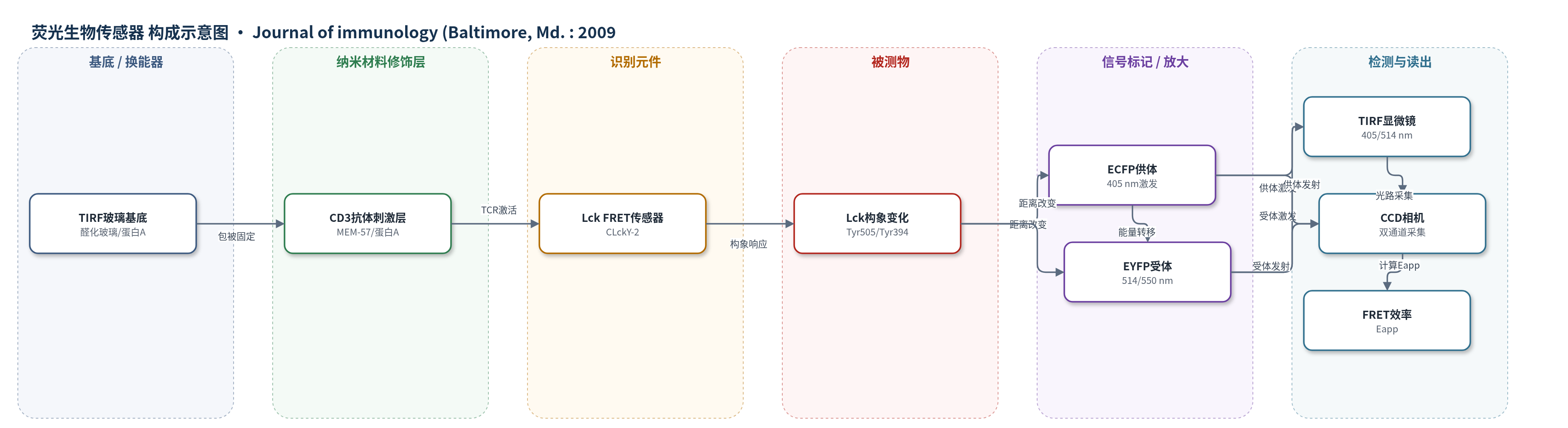
点击图片查看大图 · 依据论文自动绘制
传感器类型
荧光生物传感器
检测对象
Lck 激酶构象(Lck conformation,Tyr505/Tyr394 磷酸化相关);样品基质:Jurkat T 细胞(活细胞膜)
检测原理
该传感器以完整人源 Lck 激酶骨架为识别元件,在 SH3-SH2 连接区插入 ECFP,在 C 端 Tyr505 后融合 EYFP。Lck 的 Tyr505 去磷酸化或 Tyr394 磷酸化/突变会改变 SH2、SH3 与催化结构域之间的相对位置,从而改变 ECFP 与 EYFP 的空间距离。FRET 效率对供体-受体距离高度敏感:距离缩短时 EYFP 敏化发射增强、ECFP 供体淬灭增加;距离增大时 FRET 效率降低。TIRF 显微镜用 405 nm 激发 ECFP 并采集 480/20 nm 供体发射与 550/40 nm 受体发射,用 514 nm 激发 EYFP 校正串扰,通过受体光漂白或三立方敏化发射计算表观 FRET 效率 Eapp。该过程无外源化学放大,信号直接反映 Lck 构象状态。
检测灵敏度
原文未报告LOD、线性范围、灵敏度斜率或相关系数。
效应效果
CLckY-2 在 J.CaM1.6 中重建 CD3 诱导钙流和酪氨酸磷酸化,保留生化活性。TIRF 下 E=19±5%(n=21),ECYFP 对照 25±6%(n=95)。表达量依赖、野生型竞争和单标记共表达(Eapp=4.7±1.6%,n=65)表明高 FRET 主要来自分子内转移。Y505F 使 Eapp 降至 8.7±2.4%(n=34),Y394F 升至 22.0±1.5%(n=83),YEEI 升至 20.8±1.9%(n=61),证明对 Tyr505/Tyr394 构象敏感。TCR 激活期间 FRET 无显著变化(CD3 15.6±2.0%,n=72;BSA 16.5±3.3%,n=214),支持 Lck 激活不依赖急性磷酸化/构象改变。
传感器的构成
- 识别元件:人源 Lck 激酶完整骨架(CLckY-2),保留 N 端膜定位修饰,作为构象敏感识别元件
- 供体荧光标记:增强青色荧光蛋白(ECFP),插入 SH3-SH2 连接区,作为 FRET 供体
- 受体荧光标记:增强黄色荧光蛋白(EYFP),融合于 C 端 Tyr505 后,作为 FRET 受体
- 表达系统:逆转录病毒表达载体 pBMN-Z 与 Phoenix-gp 细胞,用于在 Jurkat T 细胞中表达传感器
- 细胞基质:Jurkat T 细胞系 E6.1 与 Lck 缺陷 J.CaM1.6,提供 TCR 信号与 Lck 重建环境
- 刺激/固定表面:蛋白 A(protein A)与 CD3 单克隆抗体 MEM-57 包被玻璃片,用于 T 细胞贴附与 TCR 激活
- 成像读出系统:TIRF 显微镜(Axiovert 200)、405/514 nm 激光与 CCD 相机(CoolSNAPHQ),用于双通道荧光采集
- 校准/对照元件:ECYFP(ECFP-EYFP 融合)用于 FRET 效率校准,CLck-2/LckY 单标记 Lck 用于区分分子内/分子间 FRET
中文摘要
当前 Src 家族激酶成员 Lck 的调控模型假定激酶骨架结构紧缩与催化活性严格相关。关键调控酪氨酸 505 磷酸化后与同一分子上的 Src 同源 2 结构域相互作用,有效抑制酪氨酸激酶活性。TCR 结合后 Tyr505 去磷酸化被认为导致激酶结构展开和活性增强。由于缺乏提供构象变化时空分辨的工具,迄今无法在活细胞中研究 Lck 构象-活性关系。我们利用完整激酶骨架设计了具有生化活性的构象敏感 Förster 共振能量转移(FRET)生物传感器,用于人源 Lck。Jurkat 细胞活细胞成像表明该传感器符合 Src 家族激酶文献报道:Tyr505 突变为 Phe 使 Lck 传感器结构打开,而将自磷酸化位点 Tyr394 突变为 Phe 使分子紧缩。高亲和 YEEI 尾突变体的紧密结构表明,在稳态条件下大多数 Lck 分子处于平均构象。尽管 T 细胞激活正常启动,我们未能在 T 细胞激活过程中检测到 Lck 生物传感器构象状态变化。结合生化数据,我们得出结论:T 细胞激活期间,Lck 可受非常细微的调控机制作用,而不需要 Tyr505 和 Tyr394 磷酸化及构象的急性改变。
英文摘要
The current model for regulation of the Src family kinase member Lck postulates a strict correlation between structural condensation of the kinase backbone and catalytic activity. The key regulatory tyrosine 505, when phosphorylated, interacts with the Src homology 2 domain on the same molecule, effectively suppressing tyrosine kinase activity. Dephosphorylation of Tyr(505) upon TCR engagement is supposed to lead to unfolding of the kinase structure and enhanced kinase activity. Studies on the conformation-activity relationship of Lck in living cells have not been possible to date because of the lack of tools providing spatiotemporal resolution of conformational changes. We designed a biochemically active, conformation-sensitive Förster resonance energy transfer biosensor of human Lck using the complete kinase backbone. Live cell imaging in Jurkat cells demonstrated that our biosensor performed according to Src family kinase literature. A Tyr(505) to Phe mutation opened the structure of the Lck sensor, while changing the autophosphorylation site Tyr(394) to Phe condensed the molecule. The tightly packed structure of a high-affinity YEEI tail mutant showed that under steady-state conditions the bulk of Lck molecules exist in a mean conformational configuration. Although T cell activation commenced normally, we could not detect a change in the conformational status of our Lck biosensor during T cell activation. Together with biochemical data we conclude that during T cell activation, Lck is accessible to very subtle regulatory mechanisms without the need for acute changes in Tyr(505) and Tyr(394) phosphorylation and conformational alterations.