组成图示
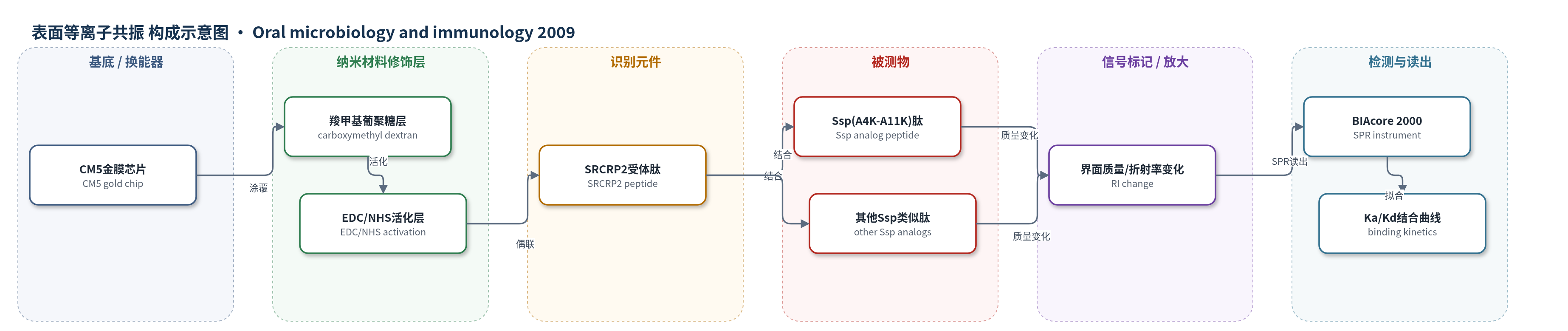
点击图片查看大图 · 依据论文自动绘制
传感器类型
表面等离子共振(SPR)生物传感器
检测对象
Ssp 类似肽(Ssp analog peptides,如 Ssp(A4K–A11K)),样品基质为 HBS-EP 缓冲液;另以唾液包被羟基磷灰石(sHA)为基质检测 S. mutans MT8148 粘附抑制。
检测原理
BIAcore CM5 芯片表面经 EDC/NHS 活化后,将唾液凝集素受体肽 SRCRP2 共价固定于羧甲基葡聚糖金膜表面。含不同赖氨酸替换的 Ssp 类似肽在 HBS-EP 缓冲液中流过芯片,与固定化 SRCRP2 发生特异性结合。结合使金膜界面局部质量与折射率改变,SPR 共振角发生偏移,仪器记录结合-解离曲线并拟合 Ka 与 Kd。Ssp(A4K–A11K) 中 4K 与 11K 的正电荷间距和方向与 SRCRP2 中 Q/E 负电荷区匹配,低离子强度下静电作用增强,因此结合响应更高。该体系无酶或核酸放大,信号直接来自肽-受体结合质量变化。
检测灵敏度
未报告 LOD、线性范围、灵敏度斜率或相关系数。
效应效果
BIAcore 实验以 4 次独立测定报告均值±标准差,pH 实验为 6 次。Ssp(A4K–A11K) 对 SRCRP2 的 Ka 为 7.6±1.1×10^3 L/m,显著高于 Ssp(A11K) 的 3.1±1.2×10^3 L/m,也高于 Q3K、A7K、A8K 类似肽;低离子强度下结合增强。双肽 Ssp(A4K–A11K)2 的 Ka/Kd 与单肽相近,提示结合依赖单受体-配体相互作用。对照共识序列肽和 PAc(316–334) 无显著结合。唾液包被羟基磷灰石实验中,0.625 µM Ssp(A11K) 及 0.5–0.75 µM Ssp(A4K–A11K) 显著抑制 S. mutans MT8148 粘附,荧光肽实验显示抑制约 50%。作者认为其可作抗粘附候选物用于龋病预防。
传感器的构成
- 基底/换能器:CM5 传感器芯片(carboxymethylated dextran-coated gold surface),提供 SPR 金膜与羧甲基葡聚糖界面
- 活化层:EDC/NHS(N-ethyl-N'-(3-diethylaminopropyl) carbodiimide/N-hydroxysuccinimide)活化羧基,生成 NHS 酯用于共价偶联
- 识别元件:SRCRP2 肽(QGRVEVLYRGSWGTVC,唾液凝集素 gp-340/DMBT1 的 SRCR 结构域肽2),固定于芯片表面捕获 Ssp 肽
- 封闭/失活层:乙醇胺盐酸盐(ethanolamine hydrochloride)封闭残余 NHS 酯,降低非特异结合
- 再生液:甘氨酸-NaOH(glycine-NaOH, pH 9.5)用于结合后再生芯片表面
- 流动相缓冲液:HBS-EP(HEPES、NaCl、EDTA、Surfactant P20),维持肽结合并优化 SPR 响应
中文摘要
戈登链球菌(Streptococcus gordonii)与牙面唾液膜相互作用,在牙菌斑生物膜形成中起重要作用。已有报道显示,SspA/SspB 的 Ssp 类似肽 A11K(第11位丙氨酸替换为赖氨酸,序列 DYQAKLAAYQAEL)可增强与唾液凝集素(gp-340/DMBT1)肽 SRCRP2 的结合。为明确赖氨酸在该结合中的作用,本研究采用 BIAcore 生物传感器检测不同赖氨酸替换 Ssp 类似肽与 SRCRP2 的结合。合成六种在表面位置引入正电荷的 Ssp 类似肽。结果显示,Ssp(A4K–A11K) 肽的结合活性显著高于其他类似肽,且在低离子强度条件下结合增强。化学计算图形分析表明,Ssp(A4K–A11K) 中 4K 与 11K 间距为 1.24±0.02 nm,接近 SRCRP2 中 Q 与 E 负电荷区间距 1.19±0.00 nm;其 1D–11K–4K 分子角小于其他肽。Ssp(A4K–A11K) 对变形链球菌结合唾液包被羟基磷灰石的抑制活性高于 A11K 肽。结论:赖氨酸定位对 Ssp 肽与 SRCRP2 结合及抑制变形链球菌牙面粘附具有重要意义。
英文摘要
INTRODUCTION: Streptococcus gordonii interacts with the salivary pellicle on the tooth surface and plays an important role in dental biofilm formation. Reports show that the analog Ssp peptide (A11K; alanine to lysine at position 11 in the arranged sequence, (1)DYQAKLAAYQAEL(13)) of SspA and SspB of S. gordonii increased binding to the salivary agglutinin (gp-340/DMBT1) peptide (scavenger receptor cysteine-rich domain 2: SRCRP2). To determine the role of lysine in the binding of the Ssp(A11K) peptide to SRCRP2, we investigated whether an additional substitution by lysine influenced the binding of Ssp(A11K) peptide to SRCRP2 using a BIAcore biosensor assay.
METHODS: Six analogs of the Ssp peptide with positive charges in surface positions on the structure were synthesized using substitution at various positions.
RESULTS: The binding activity of analog Ssp(A4K-A11K) peptide was significantly higher than the other Ssp analogs. The binding activity rose under low ionic strength conditions. The distance between positively charged amino acids in the Ssp(A4K-A11K) peptide between 4K and 11K was 1.24 +/- 0.02 nm and was close to the distance (1.19 +/- 0.00 nm) between Q and E, presenting a negative charged area, on SRCRP2 using chemical computing graphic analysis. The molecular angle connecting 1D-11K-4K in the Ssp(A4K-A11K) peptide secondary structure was smaller than the other peptide angles (1D-11K-XK). The Ssp(A4K-A11K) peptide showed higher inhibiting activity for Streptococcus mutans binding to saliva-coated hydroxyapatite than the (A11K) peptide.
CONCLUSION: The positioning of lysine is important for binding between Ssp peptide and SRCRP2, and the inhibiting effect on S. mutans binding to the tooth surface.