组成图示
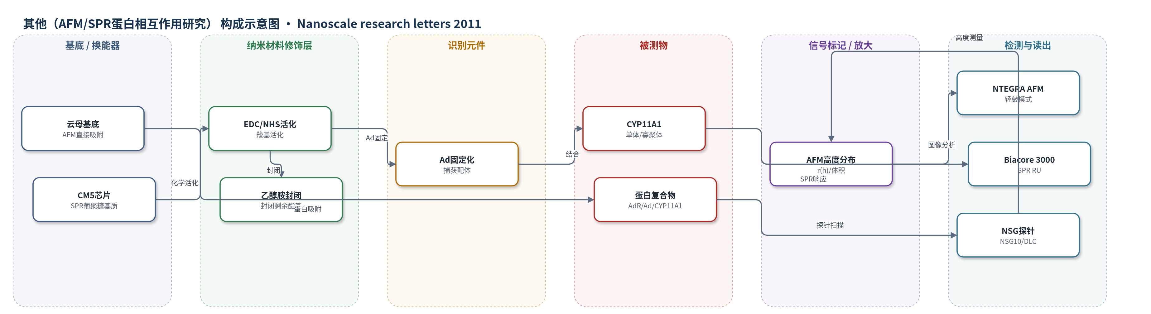
点击图片查看大图 · 依据论文自动绘制
传感器类型
其他(AFM/SPR蛋白相互作用研究)
检测对象
细胞色素CYP11A1(CYP11A1/P450scc)、肾上腺odoxin(Ad)、肾上腺odoxin还原酶(AdR)及其二元/三元复合物;样品基质为50 mM磷酸钾缓冲液(pH 7.4,含Emulgen 913)蛋白溶液。
检测原理
本文采用两种检测机制。AFM将CYP11A1、Ad、AdR及其复合物直接吸附于云母表面,轻敲模式探针扫描时,蛋白或复合物使表面局部高度增加;单体与二元/三元复合物因高度不同,在高度分布r(h)中形成不同峰,从而区分结合状态。SPR部分将Ad经EDC/NHS共价固定于CM5芯片,乙醇胺封闭后,CYP11A1流经芯片与Ad结合,界面质量与折射率变化引起表面等离子体共振响应单位(RU)变化;结合量随CYP11A1浓度增加,按1:1 Langmuir模型拟合得到kon、koff和KD。原文未使用HCR、RCA等信号放大策略。
检测灵敏度
原文未报告LOD、线性范围、灵敏度斜率或相关系数。
效应效果
AFM能区分单体与复合物:CYP11A1单体化程度达82±4%,AdR/Ad、AdR/CYP11A1和三元复合物在差分布中分别占51±8%、35±7%和12±4%。单体化后CYP11A1仍具催化活性,Vmax=0.48±0.02 nmol/min/nmol,KM=0.32±0.06 M,与寡聚体Vmax=0.51±0.04 nmol/min/nmol、KM=0.47±0.15 M无显著差异。SPR测得Ad/CYP11A1mon的kon=(290±30)×10^3 M^-1 s^-1、koff=0.05±0.005 s^-1、KD=0.17±0.015 μM,与寡聚体KD=0.21±0.02 μM接近。作者认为AFM适合研究含膜蛋白的复合物形成。
传感器的构成
- 基底/换能器:云母片(mica)作为AFM直接吸附基底,提供平整表面用于蛋白成像。
- 探针/换能元件:NSG10和NSG01_DLC微探针(NT-MDT),用于轻敲模式AFM高度/体积测量。
- 样品层:CYP11A1、Ad、AdR及其二元/三元复合物,直接吸附于云母表面形成待测对象。
- 辅助SPR基底:Biacore CM5芯片(葡聚糖基质),用于Ad/CYP11A1结合检测。
- 化学活化/封闭层:EDC/NHS活化羧基,乙醇胺封闭剩余酯基,形成固定化界面。
- 识别元件:Ad共价固定于CM5葡聚糖基质,作为捕获CYP11A1的配体。
- 被测物:CYP11A1单体/寡聚体溶液,流经芯片产生SPR响应。
- 读出系统:Biacore 3000光学生物传感器,输出响应单位(RU)并拟合kon/koff/KD。
中文摘要
本文采用原子力显微镜(AFM)和光子相关光谱(PCS)监测无磷脂条件下细胞色素CYP11A1在溶液中的单体化过程。结果表明,100 μM CYP11A1与12% Emulgen 913在50 mM KP、pH 7.4中22°C孵育10 min,可使血红素蛋白聚集体解离为单体,单体化程度为(82±4)%,且单体化后CYP11A1仍保持功能活性。AFM用于检测和可视化孤立蛋白以及CYP11A1依赖性类固醇羟化系统组分之间形成的复合物。Ad和AdR在溶液中均以单体形式存在。AFM测得单体AdR、Ad和CYP11A1的典型高度分别为1.6±0.2 nm、1.0±0.2 nm和1.8±0.2 nm;二元复合物Ad/AdR和AdR/CYP11A1mon的高度分别为2.2±0.2 nm和2.8±0.2 nm。基于光学生物传感器数据对Ad/CYP11A1mon复合物形成反应进行了动力学表征。此外,AFM还检测到典型高度为4±1 nm的三元复合物AdR/Ad/CYP11A1。
英文摘要
Atomic force microscopy (AFM) and photon correlation spectroscopy (PCS) were used for monitoring of the procedure for cytochrome CYP11A1 monomerization in solution without phospholipids. It was shown that the incubation of 100 μM CYP11A1 with 12% Emulgen 913 in 50 mM KP, pH 7.4, for 10 min at T = 22°C leads to dissociation of hemoprotein aggregates to monomers with the monomerization degree of (82 ± 4)%. Following the monomerization procedure, CYP11A1 remained functionally active. AFM was employed to detect and visualize the isolated proteins as well as complexes formed between the components of the cytochrome CYP11A1-dependent steroid hydroxylase system. Both Ad and AdR were present in solution as monomers. The typical heights of the monomeric AdR, Ad and CYP11A1 images were measured by AFM and were found to correspond to the sizes 1.6 ± 0.2 nm, 1.0 ± 0.2 nm and 1.8 ± 0.2 nm, respectively. The binary Ad/AdR and AdR/CYP11A1mon complexes with the heights 2.2 ± 0.2 nm and 2.8 ± 0.2 nm, respectively, were registered by use of AFM. The Ad/CYP11A1mon complex formation reaction was kinetically characterized based on optical biosensor data. In addition, the ternary AdR/Ad/CYP11A1 complexes with a typical height of 4 ± 1 nm were AFM registered.