组成图示
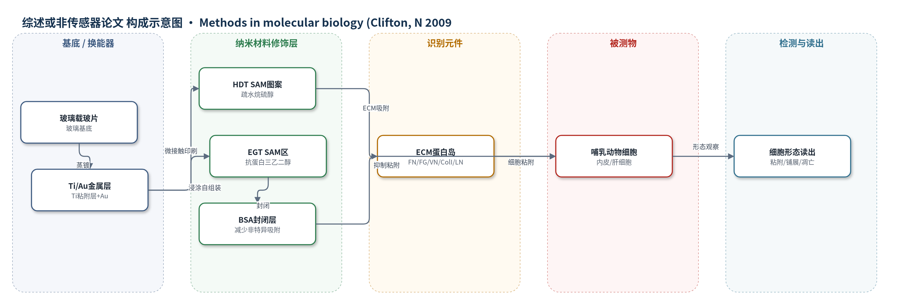
点击图片查看大图 · 依据论文自动绘制
传感器类型
综述或非传感器论文
检测对象
无明确检测对象(非传感检测);涉及细胞外基质蛋白(extracellular matrix proteins, ECM)和哺乳动物细胞(mammalian cells),样品基质为 PBS 或细胞培养液。
检测原理
该方法不产生电化学或光学传感信号,而是通过表面化学图案化控制细胞粘附。首先,微接触印刷将十六烷硫醇(HDT)转移到金表面,形成疏水自组装单分子层图案;剩余金面用三乙二醇硫醇(EGT)处理,形成抗蛋白吸附区。随后,细胞外基质(ECM)蛋白从 PBS 中选择性吸附到 HDT 疏水区,而 EGT 区排斥蛋白,形成 ECM 粘附岛。BSA 封闭后,细胞通过细胞–ECM 相互作用仅能在图案区粘附、铺展;非粘附区抑制细胞附着。细胞铺展状态改变细胞形状与力学信号,从而在生长与凋亡程序之间切换。读出为细胞形态、位置、存活或凋亡状态,而非浓度依赖的传感信号。
检测灵敏度
—
效应效果
该方法可制备特征尺寸大于 500 nm、特殊情况下约 200 nm 的粘附/非粘附图案。HDT 区可选择性吸附纤连蛋白、纤维蛋白原、玻连蛋白、I 型胶原和层粘连蛋白等 ECM 蛋白,使牛毛细血管内皮细胞和大鼠肝细胞仅粘附于图案区;EGT 区抗蛋白吸附,减少非特异粘附。金基底蒸镀后 4–5 周可能出现斑驳,立即进行微接触印刷可稳定约 3 个月;烷硫醇乙醇溶液保存超过 3 个月会氧化生成二硫化物,其自组装速度比硫醇慢约 75 倍。作者认为该技术可用于细胞培养、组织工程和细胞生物传感器开发,通过几何与粘附微环境调控细胞形状、位置和功能。
传感器的构成
- 基底:玻璃载玻片(Fisher no.2),提供平整支撑。
- 金属粘附层:Ti(15 Å),增强金与玻璃结合。
- 金属基底:Au(115 Å),烷硫醇自组装基底。
- 疏水SAM图案:十六烷硫醇(HDT),微接触印刷形成ECM蛋白吸附岛。
- 抗蛋白SAM:三乙二醇硫醇(EGT, HS(CH2)11O(CH2CH2O)2CH2CH2OH),覆盖剩余金面,抗蛋白吸附和细胞粘附。
- ECM蛋白层:纤连蛋白(FN)、纤维蛋白原(FG)、玻连蛋白(VN)、I型胶原(Col I)、层粘连蛋白(LN)等,选择性吸附于HDT区,介导细胞粘附。
- 封闭剂:牛血清白蛋白(BSA),封闭非图案区,减少非特异吸附。
- 细胞层:哺乳动物细胞(如牛毛细血管内皮细胞、大鼠肝细胞),仅粘附并铺展于ECM图案区。
中文摘要
本文介绍一种利用微接触印刷(μCP)将烷硫醇转移到金表面、构建自组装单分子层(SAMs)图案化基底的方法。该方法在玻璃载玻片上蒸镀钛和金,用光刻制作 PDMS 印章,将十六烷硫醇(HDT)印刷到金表面形成疏水图案,再将剩余金面暴露于三乙二醇硫醇(EGT)形成抗蛋白吸附区。随后,细胞外基质(ECM)蛋白选择性吸附于 HDT 图案区,而 EGT 区排斥蛋白,从而形成 ECM 粘附岛与非粘附区。哺乳动物细胞仅能粘附并铺展于 ECM 图案区。作者利用该技术证明,在饱和生长因子存在下,通过促进或阻止细胞铺展,可将哺乳动物细胞在生长与凋亡程序之间切换。结果表明,局部细胞–ECM 相互作用和细胞形状可调控细胞生理,微图案化技术有助于理解 ECM 与细胞形状对细胞功能的影响,并可用于细胞培养、组织工程和细胞生物传感器开发。
英文摘要
We present a method that uses microcontact printing of alkanethiols on gold to generate patterned substrates presenting "islands" of extracellular matrix (ECM) surrounded by nonadhesive regions such that single cells attach and spread only on the adhesive regions. We have used this micropatterning technology to demonstrate that mammalian cells can be switched between growth and apoptosis programs in the presence of saturating concentrations of growth factors by either promoting or preventing cell spreading (Science 276:1425-1428, 1997). From the perspective of fundamental cell biology, these results suggested that the local differentials in growth and viability that are critical for the formation of complex tissue patterns may be generated by local changes in cell-ECM interactions. In the context of cell culture technologies, such as bioreactors and cellular engineering applications, the regulation of cell function by cell shape indicates that the adhesive microenvironment around cells can be carefully optimized by patterning a substrate in addition to using soluble factors (Biotech. Prog. 14:356-363, 1998). Micropatterning technology is playing a central role both in our understanding how ECM and cell shape regulate cell physiology and in facilitating the development of cellular biosensor and tissue engineering applications (Science 264:696-698, 1994; J. Neurosci. Res. 13:213-20, 1985; Biotech. Bioeng. 43:792-800, 1994).