组成图示
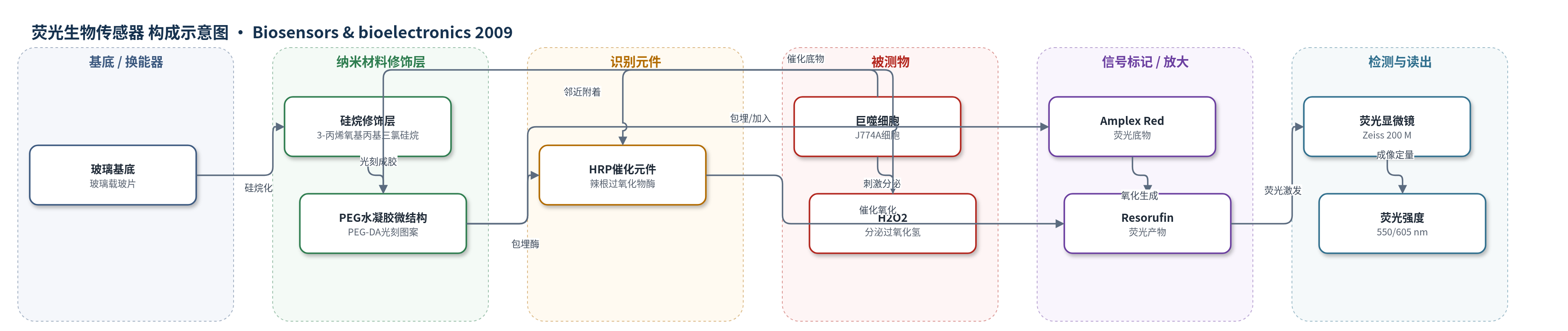
点击图片查看大图 · 依据论文自动绘制
传感器类型
荧光生物传感器
检测对象
过氧化氢(H2O2),样品基质为巨噬细胞(J774A)培养上清/培养基(DMEM)
检测原理
巨噬细胞经 PMA 刺激后发生氧化爆发,向培养基分泌 H2O2。H2O2 扩散进入邻近的 PEG-DA 水凝胶微结构,与包埋其中的 HRP 接触。HRP 以 H2O2 为底物催化氧化 Amplex Red,生成荧光产物 Resorufin;Amplex Red 可预先包埋于水凝胶中,也可在检测时加入培养基。荧光强度随 H2O2 浓度升高而增强,校准曲线在 0–20 μM 范围内呈线性,高浓度区因 HRP 抑制或 Resorufin 进一步氧化而斜率降低。系统通过荧光显微镜在 550 nm 激发、605 nm 发射下采集图像,并用 1 μM H2O2 参考点归一化,再对照校准曲线定量细胞分泌的 H2O2。
检测灵敏度
线性范围: 0–20 μM H2O2
效应效果
该传感器在微图案化表面保持抗污性,巨噬细胞和成纤维细胞仅附着于暴露玻璃区,不粘附含 HRP 的 PEG 水凝胶;无酶 PEG 微结构作为负控无荧光,仅 HRP 凝胶响应 H2O2,显示空间选择性。PMA 刺激巨噬细胞后,水凝胶荧光较未刺激细胞增加超过 2 倍;三次独立实验中刺激/未刺激信号比稳定为 2.1±0.1,表明方案可重复。作者用标准 HRP/Amplex Red 微孔板荧光法验证了细胞 H2O2 分泌。1 h 与 3 h PMA 刺激对应 H2O2 浓度约 1 μM 与 0.55 μM,提示氧化爆发具有短暂性。系统无需收集培养基离线分析,可在培养皿原位监测小群体细胞代谢,未来可扩展至单细胞阵列和多代谢物检测。
传感器的构成
- 基底:玻璃载玻片(glass slides),经硅烷(3-acryloxypropyl trichlorosilane)修饰以固定 PEG 水凝胶
- 修饰层:PEG-DA 水凝胶微结构(poly(ethylene glycol) diacrylate, MW 575,含 2% v/v 光引发剂 2-hydroxy-2-methyl-propiophenone),光刻图案化,抗污并承载酶
- 识别/催化元件:HRP(horseradish peroxidase),包埋于 PEG-DA 水凝胶中,催化 H2O2 氧化 Amplex Red
- 信号标记物:Amplex Red,荧光底物,可包埋于水凝胶或加入培养基,被 HRP/H2O2 氧化为荧光 Resorufin
- 细胞附着区:暴露玻璃区,引导巨噬细胞附着于传感元件旁,避免细胞覆盖传感结构
中文摘要
传感元件与小群体细胞的整合是细胞培养与分析微型化的关键步骤。本文报道了一种用于局部检测受刺激巨噬细胞分泌过氧化氢(H2O2)的光学酶基生物传感器。通过光刻图案化含辣根过氧化物酶(HRP)的聚乙二醇(PEG)水凝胶微结构,在玻璃表面构建传感结构。包埋酶的水凝胶微图案不支持蛋白或细胞沉积,并能引导巨噬细胞附着于传感元件旁。Amplex Red 是一种在 H2O2 和 HRP 存在下产生荧光的有机分子,可随酶分子包埋于水凝胶元件内,或在细胞激活时加入细胞培养基中。巨噬细胞经有丝原刺激后产生 H2O2,使含 HRP 的水凝胶微结构出现荧光,且荧光强度与分析物浓度密切相关。该新型集成传感元件的细胞培养系统未来可通过引入额外生物识别元件,实现细胞位点多代谢物检测。
英文摘要
The integration of sensing elements with small groups of cells is a critical step towards miniaturization of cell cultivation and analysis. This paper describes the development of an optical, enzyme-based biosensor for local detection of hydrogen peroxide (H(2)O(2)) secreted by stimulated macrophages. Photolithographic patterning of horseradish peroxidase (HRP)-containing poly (ethylene glycol) (PEG) hydrogel microstructures was used to create sensing structures on the glass surface. Importantly, enzyme-entrapping hydrogel micropatterns did not support protein or cell deposition and allowed to guide attachment of macrophages next to the sensing elements. Amplex Red, an organic molecule that becomes fluorescent in the presence of H(2)O(2) and HRP, was either immobilized inside hydrogel elements alongside enzyme molecules or added into the cell culture media during cell activation. The production of H(2)O(2) after mitogenic stimulation of macrophages resulted in appearance of fluorescence in the HRP-containing hydrogel microstructures, with fluorescence intensity being a strong function of analyte concentration. The novel cell culture system with integrated sensing elements described here may be enhanced in the future by incorporating additional biorecognition elements to enable multi-metabolite detection at the site of a cell.