组成图示
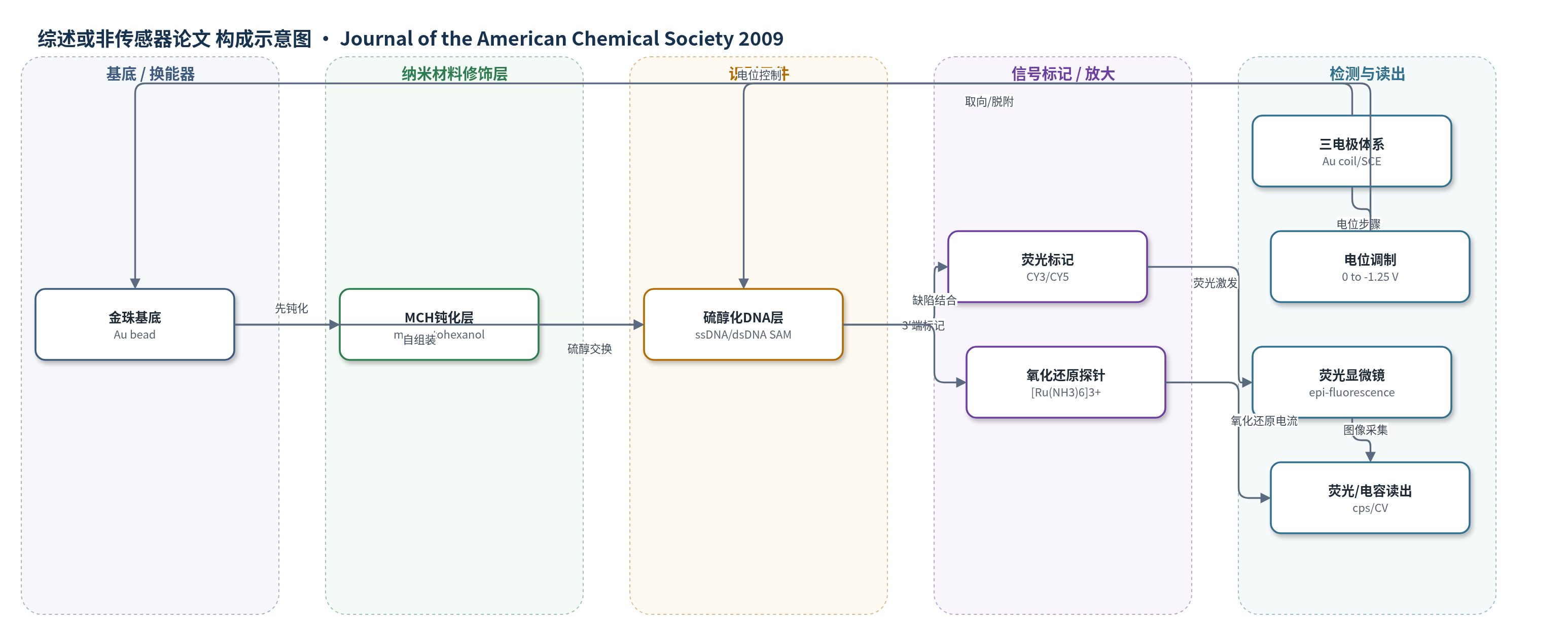
点击图片查看大图 · 依据论文自动绘制
传感器类型
综述或非传感器论文
检测对象
无明确分析物;研究对象为硫醇化 DNA 自组装单分子层(DNA SAM)修饰金表面,样品基质为 10 mM Tris/10 mM NaBF4 电解液。
检测原理
该研究并非以特定分析物浓度产生信号,而是表征 DNA 修饰金电极的界面行为。硫醇化 DNA 通过 5′-硫醇端与 Au 表面自组装,3′端 CY3/CY5 荧光基团距离金表面越近,荧光受金属猝灭越强。改变电极电位时,Au 表面电荷变化使 DNA 链发生取向改变或静电排斥,荧光强度随之变化;负电位下硫醇键被还原,DNA 从表面脱附并远离金表面,猝灭减弱,荧光增强,同时微分电容上升。[Ru(NH3)6]3+ 通过 SAM 缺陷接近电极发生氧化还原,其峰电流用于估算 DNA 表面密度。若将该界面用于杂交传感,目标 DNA 结合会改变链取向、堆积和荧光调制幅度,从而实现识别事件到荧光/电化学信号的转换。
检测灵敏度
原文未报告 LOD、线性范围、灵敏度斜率或相关系数(R^2)。
效应效果
DNA/MCH 表面存在显著异质性,强荧光热点覆盖约10–30%表面,ssDNA 比 dsDNA 更多。高表面密度 ssDNA 在 0 至 -400 mV 区间荧光调制小于2%。先 MCH 后 DNA 的制备使表面密度降至 1.1×10^10 和 3.7×10^10 strands/cm2,较原方法低约100–1000倍,热点明显减少;MCH/ssDNA 电位调制显著,MCH/dsDNA 平均调制约20%,区域变化约10–25%。还原脱附证明表面完全修饰,ssDNA/MCH 脱附始于 -775 mV,dsDNA/MCH 始于 -650 mV,MCH/DNA 始于 -625 mV。还观察到 3–5 µm 荧光颗粒移动,速度 0–10 µm/s,支持物理吸附 DNA 聚集体。作者认为该表征与制备策略有助于改善 DNA 传感器表面质量。
传感器的构成
- 基底/换能器电极:金珠(Au bead,约1–2.5 mm),经piranha清洗和火焰退火,用作工作电极。
- 识别元件:5′-己硫醇修饰单链/双链 DNA 探针(ssDNA/dsDNA,LN1/LN5),通过硫醇-Au自组装形成DNA SAM。
- 信号标记物:3′端 CY3 或 CY5 荧光基团,用于荧光成像与电位调制。
- 封闭/钝化层:己硫醇(MCH, mercaptohexanol),填充表面并置换非特异性吸附DNA。
- 氧化还原探针:[Ru(NH3)6]3+,通过SAM缺陷结合,用于CV测定DNA表面密度。
- 电化学介质:10 mM Tris 与 10 mM NaBF4 电解液,提供离子导电与电位控制环境。
- 三电极体系:Au线圈对电极与SCE参比电极,用于CV、微分电容和电位步骤。
- 荧光读出装置:倒置荧光显微镜(Olympus IX70,U-MNG2滤光片组,SPOT RT相机),原位采集荧光图像。
中文摘要
本文结合电化学研究与荧光成像,研究金表面由单链或双链 DNA 自组装单分子层(SAM)修饰后产生的表面异质性。直接吸附硫醇化 DNA 并用己硫醇(MCH)钝化后,表面并非均匀:一部分区域形成 DNA SAM,另一部分被大尺寸 DNA 聚集体覆盖,二者荧光强度差异显著。更重要的是,荧光强度随电极电位变化呈现区域差异,大聚集体与单分子层区域的电位调制明显不同。通过负电位还原脱附并检测荧光标记 DNA,证明表面被完全修饰,但修饰程度在空间上不均匀。研究结果对基于 DNA SAM 的生物传感器和生物芯片开发具有启示。作者进一步采用先 MCH 成膜再引入硫醇化 DNA 的替代方法,获得聚集体更少、荧光电位调制更显著的表面,但 DNA 表面密度显著降低,约为最大理论覆盖的 1%。该工作强调使用电化学原位荧光显微镜等非平均化成像手段研究传感器表面,以改善 DNA 传感器界面质量。
英文摘要
The creation of gold surfaces modified by single- or double-stranded DNA self-assembled monolayers (SAMs) is shown to produce heterogeneous surface packing densities through the use of electrochemical studies coupled with fluorescence imaging. The modified surfaces created by direct adsorption of thiolate DNA [followed by passivation with mecaptohexanol (MCH)] resulted in regions covered by a monolayer of DNA SAM and other regions that were coated by large particles of DNA. The difference in fluorescence intensity measured from these regions was dramatic. More importantly, a regional variance in fluorescence intensity in response to electrochemical potential was observed: the large aggregates showing a significantly different modulation of fluorescence intensity than the monolayer-coated regions. Electrochemical desorption and detection of the fluorescently tagged DNA provided clear evidence of a complete surface modification. These studies have implications for biosensor/biochip development using DNA SAMs. A modification in the method used to produce the DNA SAMs resulted in a significantly different surface with much fewer aggregates and more significant electromodulation of the fluorescence intensity, though at much lower DNA surface density (ca. 1% of maximum theoretical coverage). This method for forming the modified surfaces has clear advantages over the currently accepted practice and emphasizes the importance of studying the nonaveraged nature of the sensor surface using in situ imaging tools like electrofluorescence microscopy.