组成图示
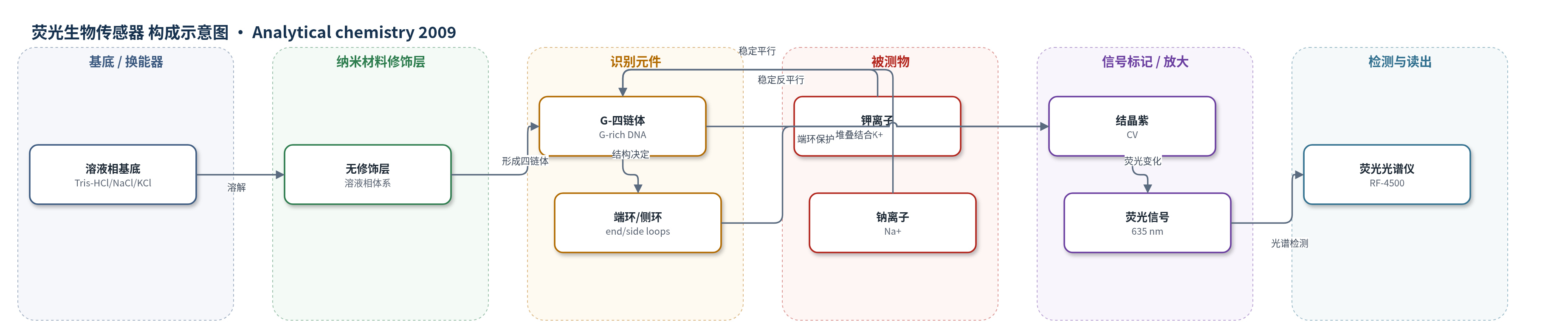
点击图片查看大图 · 依据论文自动绘制
传感器类型
荧光生物传感器
检测对象
G-四链体结构(G-quadruplex structures)、钾离子(potassium ion, K+);样品基质:25 mM Tris-HCl 缓冲液(含 NaCl/KCl)
检测原理
该传感器为溶液相荧光体系。富含 G 的寡核苷酸在 K+ 或 Na+ 存在下折叠为 G-四链体,其拓扑结构由离子决定:K+ 倾向于稳定平行结构,Na+ 倾向于稳定反平行或混合结构。结晶紫(CV)以 2:1 化学计量堆叠到 G-四链体两端外部 G-四分体上。当 G-四链体为反平行构型时,端环将结合态 CV 与溶剂隔离,降低非辐射猝灭,使 CV 荧光显著增强;平行构型只有侧环,保护不足,荧光较弱。因此,CV 荧光强度可反映 G-四链体结构。在 Na+ 浓度达到饱和(如 600 mM)后,加入 K+ 会促使 G-四链体由反平行/混合结构向平行结构转变,CV 荧光随之下降。通过监测 635 nm 处荧光强度变化,即可检测 K+ 浓度并监测结构互变。
检测灵敏度
原文未报告 LOD、线性范围、灵敏度斜率或相关系数。
效应效果
该方法对 Na+ 选择性高:200 mM Na+ 变化仅相当于 0.3 mM K+ 的荧光变化;800 mM Na+ 下 2.0 mM K+ 与 600 mM Na+ 下 1.7 mM K+ 信号相当。CV 与 Oxy12、Oxy28、Oxy32 结合化学计量均为 2:1,Ka 分别为 0.0066±0.0003、0.019±0.003、0.012±0.001 µM^-2;与 Hum12、Hum21、Hum24 的 Ka 分别为 0.0033±0.0008、0.036±0.008、0.059±0.018 µM^-2。竞争透析表明结合偏好不随离子条件显著改变,荧光差异主要来自端环保护。方法简单、廉价,无需标记 DNA,可耐受大量 Na+,适用于 K+ 检测和 G-四链体结构监测。
传感器的构成
- 基底/换能器:无固定基底或电极,溶液相荧光检测体系,CV 与 DNA 在缓冲液中混合
- 纳米材料修饰层:无纳米材料修饰层,直接溶液相混合
- 识别元件:富含 G 的寡核苷酸(G-rich oligonucleotides,如 Oxy12、Oxy28、Oxy32、Hum12、Hum21、Hum24、d(G3TiG3TjG3TkG3)),形成 G-四链体
- 结构识别元件:G-四链体端环(end loops)与侧环(side loops),端环保护结合态 CV,侧环保护弱
- 信号标记物:结晶紫(CV),三苯甲烷染料,与 G-四链体外部 G-四分体堆叠,荧光增强
- 离子识别/调节层:K+ 或 Na+ 离子,调控 G-四链体平行/反平行结构互变,用于 K+ 检测
- 缓冲介质:25 mM Tris-HCl(pH 7.0)含 NaCl/KCl,维持 pH、离子强度和四链体形成
- 读出层:荧光光谱仪(Hitachi RF-4500),激发 580 nm,发射 610–700 nm,监测 635 nm 荧光强度
中文摘要
基因组 DNA 中富含鸟嘌呤的序列可形成 G-四链体,其生物学功能可能依赖结构。开发能区分不同类型 G-四链体的敏感结构探针备受关注。结晶紫(CV)是一种三苯甲烷染料,可堆叠到 G-四链体两个外部 G-四分体上,此前已被用于区分 G-四链体与双链/单链 DNA。本文研究了 CV 区分平行与反平行 G-四链体结构的能力。CV 与反平行 G-四链体结合后,由于端环将结合态 CV 与溶剂隔离,其荧光强度显著升高;而平行 G-四链体中的侧环不能提供类似保护,因此 CV/G-四链体混合物的荧光明显弱于反平行结构。据此,CV 可发展为敏感荧光生物传感器,用于区分反平行与平行 G-四链体并监测 G-四链体结构互变。此外,由于某些富含 G 的 DNA 序列在 Na+ 或 K+ 条件下可采取不同 G-四链体结构,作者开发了一种新颖、廉价、简单的 K+ 离子检测方法。该方法对 Na+ 具有高选择性,200 mM Na+ 浓度变化仅引起与 0.3 mM K+ 相近的荧光信号变化。
英文摘要
G-rich sequences with the potential for quadruplex formation are common in genomic DNA. Considering that the biological functions of G-quadruplexes may well depend on their structures, the development of a sensitive structural probe for distinguishing different types of quadruplexes has received great attention. Crystal violet (CV) is a triphenylmethane dye, which can stack onto the two external G-quartets of a G-quadruplex. The ability of CV to discriminate G-quadruplexes from duplex and single-stranded DNAs has been reported by us. Herein, the ability of CV to discriminate parallel from antiparallel structures of a G-quadruplex was studied. The binding of CV to an antiparallel G-quadruplex can make its fluorescence intensity increase to a high level because of the protection of bound CV from the solvent by quadruplex end loops. The presence of side loops in parallel G-quadruplexes cannot provide bound CV such protection, causing the fluorescence intensity of CV/G-quadruplex mixture to be obviously weaker when the G-quadruplex adopts a parallel structure than that when the G-quadruplex adopts an antiparallel structure. Therefore, CV can be developed as a sensitive fluorescent biosensor for the discrimination of antiparallel G-quadruplexes from parallel G-quadruplexes and for monitoring the structural interconversion of G-quadruplexes. In addition, considering that some G-rich DNA sequences can adopt different G-quadruplex structures under Na(+) or K(+) ion conditions, a novel, cheap and simple K(+) ion detection method was developed. This method displays a high K(+) ion selectivity against Na(+) ion, the change of 200 mM in Na(+) ion concentration only causes a similar fluorescent signal change to 0.3 mM K(+) ion.