组成图示
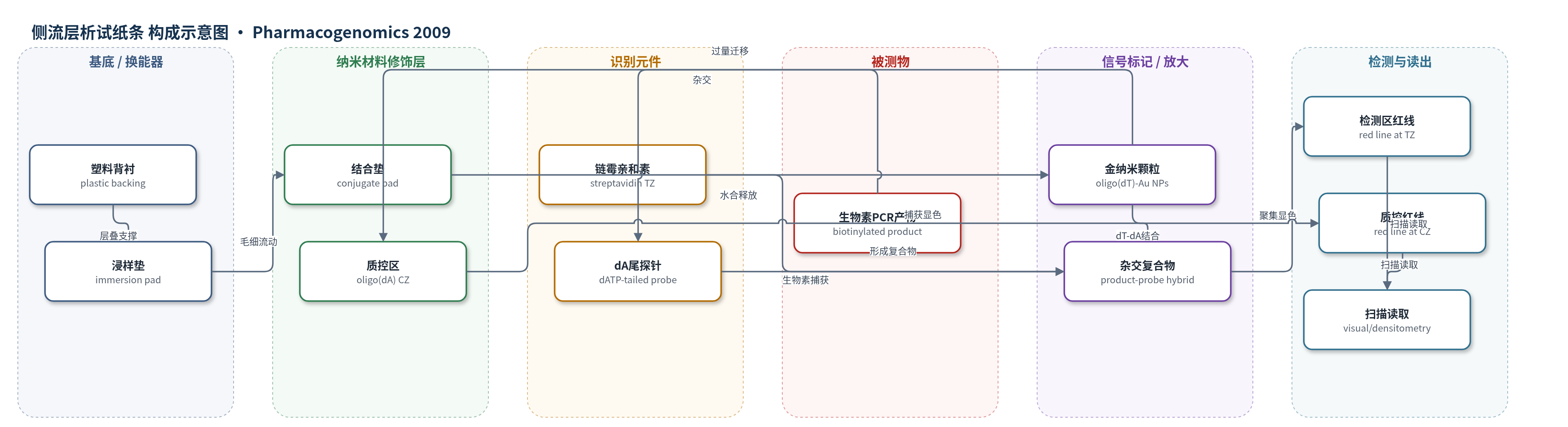
点击图片查看大图 · 依据论文自动绘制
传感器类型
侧流层析试纸条
检测对象
CYP2C19*2(c.681G>A)和CYP2D6*4(g.3465G>A)单核苷酸多态性(SNP)基因型;样品基质:全血基因组DNA
检测原理
全血基因组DNA经四引物PCR扩增:外部引物先扩增覆盖SNP的长片段;降温后3′端错配、5′端生物素标记的内部引物与外部引物配对,双向扩增产生等位基因特异性短产物。短产物与带poly(dA)尾的N/M探针杂交后点样至试纸条。缓冲液毛细迁移使结合垫中oligo(dT)-Au NPs水合;检测区链霉亲和素捕获生物素标记短产物,探针dA尾与金颗粒oligo(dT)杂交,使金颗粒聚集并在520 nm处形成红色检测线。质控区oligo(dA)捕获多余颗粒形成第二条红线。长产物无生物素而不被捕获。N/M两条试纸的红线组合判断N/N、N/M或M/M基因型,信号强度与等位基因特异性产物量相关。
检测灵敏度
可检测量: as low as 3 fmol of amplified DNA;易检量: 10 and 27 fmol of the N- and M-specific fragments, respectively;信号随靶量增加至: 40 and 109 fmol, respectively;N:M产物比范围: 1/0.03 to 1/144。
效应效果
方法对55例CYP2C19*2和49例CYP2D6*4全血样品(共208个等位基因)进行分型,结果与测序及2%琼脂糖凝胶电泳完全一致。重现性CV为:N/N样品N探针6.8%、M探针10%;N/M样品N探针11%、M探针3.5%;M/M样品N探针15%、M探针4.6%。可检测低至3 fmol扩增DNA,对N:M产物比1/0.03至1/144具有特异性。杂交温度至42°C信号稳定,探针1–2 pmol后信号恒定。试纸条10 min内显色,总耗时2–2.5 h,成本约2€/次。相比电泳,杂交提供序列确认并减少非特异条带干扰,适合小型临床实验室和即时检测。
传感器的构成
- 基底/支撑层:塑料粘胶背衬(plastic adhesive backing),承载浸样垫、结合垫、层压膜和吸收垫并维持层叠结构。
- 浸样垫:immersion pad,接收杂交液/样品并启动毛细流动。
- 结合垫:conjugation pad,预置寡聚(dT)偶联金纳米颗粒(oligo(dT)-Au NPs,40 nm),作为信号标记物。
- 层压膜检测区:laminated membrane test zone,固定链霉亲和素(streptavidin, SA),捕获生物素标记的等位基因特异性PCR短产物。
- 层压膜质控区:laminated membrane control zone,固定寡聚(dA)(oligo(dA)),捕获多余金纳米颗粒形成质控红线。
- 吸收垫:absorbent pad,吸收流动缓冲液并维持毛细迁移。
- 流动缓冲液:PBS含4%甘油和1% SDS,提供毛细流动介质并维持杂交与颗粒分散。
- 识别/捕获元件:样品中dATP尾寡核苷酸探针(N/M probe)与PCR产物杂交,链霉亲和素-生物素作用实现序列确认与捕获。
中文摘要
单核苷酸多态性(SNP)分型通常包括指数扩增、等位基因判别和产物检测,且判别多在PCR后进行。四引物PCR可在扩增过程中完成等位基因判别,但迄今主要依赖电泳检测。本文报道一种干试剂试纸条DNA生物传感器,可在数分钟内无仪器可视化检测四引物PCR产物,并应用于CYP2C19*2(c.681G>A)和CYP2D6*4(g.3465G>A)基因分型。方法中,一对外部引物扩增覆盖SNP的片段;5′端生物素标记、3′端错配的内部引物与外部引物配对,引导双向扩增产生等位基因特异性短片段。PCR产物先与带poly(dA)尾的探针短暂杂交,再点样至生物传感器并浸入缓冲液。缓冲液沿试纸条迁移时,杂交复合物被检测区链霉亲和素捕获,并与寡聚(dT)功能化金纳米颗粒结合,形成红色检测线;质控区形成第二条红线以确认传感器正常工作。作者对55例CYP2C19*2和49例CYP2D6*4样品进行分型,结果经测序和电泳确认。该方法简单、低成本,杂交提供序列确认,干试剂试纸条降低对专业人员的要求。
英文摘要
BACKGROUND: SNP-typing strategies involve an exponential amplification step, an allele discrimination reaction and detection of the products. Usually, allele discrimination is performed after amplification. Tetra-primer PCR allows allele discrimination during the amplification step, thereby avoiding additional genotyping reactions. However, to date, electrophoresis is the only method used for detection of tetra-primer PCR products. We report a dipstick test that enables visual detection of tetra-primer PCR products within minutes without instruments. The method is applied to the genotyping of CYP2C19*2 (c.681G>A) and CYP2D6*4 (g.3465G>A).
MATERIALS & METHODS: A pair of external primers amplifies a segment encompassing the SNPs. Biotinylated inner primers have a 3 -mismatch and pair off with the external primers to guide a bidirectional amplification that generates allele-specific fragments. The products are hybridized briefly with poly(dA)-tailed probes and applied to the DNA biosensor, which is then immersed in the appropriate buffer. As the buffer migrates along the biosensor, the hybrids are captured from streptavidin at the test zone and interact with oligo(dT)-functionalized gold nanoparticles leading to the formation of a red line. Another red line is formed at the control zone to indicate proper function of the sensor.
RESULTS: We genotyped 55 samples for CYP2C19*2 and 49 samples for CYP2D6*4. The accuracy of this method was confirmed by sequencing and electrophoresis.
CONCLUSIONS: The unique advantages of the proposed method are its simplicity and low cost. Contrary to electrophoresis, hybridization provides sequence confirmation of amplified fragments. The dry-reagent dipstick format minimizes the requirements for highly qualified personnel.