组成图示
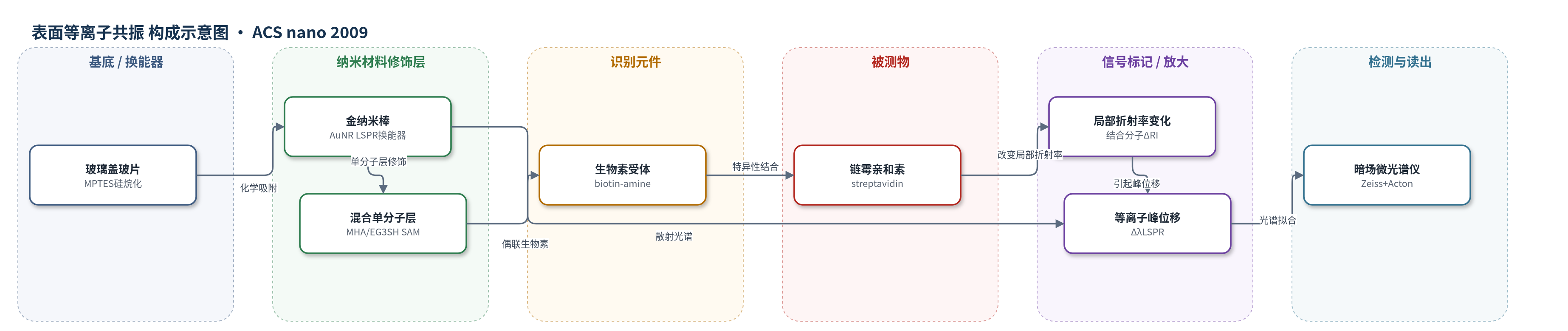
点击图片查看大图 · 依据论文自动绘制
传感器类型
表面等离子共振(SPR)生物传感器
检测对象
链霉亲和素(streptavidin);样品基质:PBS/水溶液(实验在 PBS 中孵育,光谱拟合在水/缓冲液)
检测原理
金纳米棒表面导带电子在光激发下发生局域表面等离子共振(LSPR),其散射峰波长和强度对纳米棒周围局部折射率敏感。传感器表面经 MHA/EG3SH 单分子层和生物素功能化后,链霉亲和素与生物素特异性结合,使纳米棒附近光学质量增加、局部折射率升高,导致 LSPR 峰发生红移。模型将峰位移 Δλ 与结合分子数 N 近似线性关联:Δλ 正比于 S(r)·ΔRI·N·VA/VS,其中 S(r) 为距离相关折射率灵敏度,VS 为传感体积,VA 为分子体积。暗场微光谱仪采集单个纳米棒散射光谱并拟合峰位置,当位移超过系统不确定度(约0.3 nm)即可判定结合。该过程无标记、无酶放大,灵敏度由纳米棒几何、散射截面、传感体积和检测系统噪声共同决定。
检测灵敏度
LOD: 160 pM;MDL: 18 个链霉亲和素分子;实际纳米棒平均 MDL: 20 个分子;实验最低可测浓度: 100 pM(约27个分子/纳米棒);饱和位移: 5.4 nm(约280个分子)
效应效果
实验以过量游离生物素预饱和链霉亲和素作为阴性对照,验证生物素–链霉亲和素结合的特异性;EG3SH 混合单分子层用于抑制非特异吸附。系统峰测量不确定度约0.3 nm,拟合不确定度约0.02 nm,每个浓度约测量15根纳米棒并给出95%置信区间。生物素化引起平均3.23±1.2 nm位移,100 pM链霉亲和素引起0.52 nm位移,对应约27个分子;饱和位移5.4 nm对应约280个分子。作者称160 pM为单颗粒LSPR传感器报道的最低生物分子检测浓度,并认为通过优化几何、光源、受体长度和检测稳定性,MDL可再降低约800倍,有望实现单分子结合检测。
传感器的构成
- 基底:玻璃盖玻片(glass coverslip),经 MPTES(mercaptopropyltriethoxysilane)硅烷化,用于固定金纳米棒并提供光学透明支撑
- 换能纳米材料:金纳米棒(Au nanorods,约63×25 nm),化学吸附于 MPTES 化玻璃表面,产生 LSPR 散射信号
- 自组装单分子层:MHA(原文 mercapto-hexadecanoic acid)与 EG3SH((1-mercaptoundec-11-yl)tri(ethylene glycol))混合 SAM,MHA 提供羧基用于偶联生物素,EG3SH 封闭非特异吸附并调节受体密度
- 识别元件:生物素(biotin-amine),经 EDC/NHS 活化 MHA 羧基后偶联,特异性结合链霉亲和素
- 信号标记物:无外源标记,链霉亲和素结合改变纳米棒周围局部折射率,引起 LSPR 峰波长位移
- 读出系统:暗场微光谱仪(dark-field microspectrometer,Zeiss Axiovert 200 + Acton SpectraPro 2150i),采集单个纳米棒散射光谱并拟合 LSPR 峰
中文摘要
本文提出一种用于理性设计基于单个金纳米颗粒局域表面等离子共振(LSPR)位移的无标记生物传感器的解析模型。该模型将单个等离子体纳米颗粒散射光的峰波长与其表面结合的分析物分子数相关联,并给出解析公式,以预测传感器关键性能指标,如分子检测限(MDL)和动态范围;这些指标可表示为纳米颗粒几何尺寸和检测系统参数的函数。模型可计算单个金纳米棒结合单个分子时引起的 LSPR 位移,因此 MDL 定义为系统可可靠测得的最小结合分子数,动态范围定义为单个纳米棒可检测的最大分子数。该模型可用于在实验前针对目标分析物优化 LSPR 传感器性能。作者利用该模型设计了基于生物素功能化金纳米棒的 LSPR 传感器,并预测其可获得该类传感器中最低的 MDL,即 18 个链霉亲和素分子;该预测与实验结果和估算相符。文章进一步讨论了如何利用该模型指导未来 LSPR 生物传感器的优化与单分子检测发展。
英文摘要
We present the development of an analytical model that can be used for the rational design of a biosensor based on shifts in the local surface plasmon resonance (LSPR) of individual gold nanoparticles. The model relates the peak wavelength of light scattered by an individual plasmonic nanoparticle to the number of bound analyte molecules and provides an analytical formulation that predicts relevant figures-of-merit of the sensor such as the molecular detection limit (MDL) and dynamic range as a function of nanoparticle geometry and detection system parameters. The model calculates LSPR shifts for individual molecules bound by a nanorod, so that the MDL is defined as the smallest number of bound molecules that is measurable by the system, and the dynamic range is defined as the maximum number of molecules that can be detected by a single nanorod. This model is useful because it will allow a priori design of an LSPR sensor with figures-of-merit that can be optimized for the target analyte. This model was used to design an LSPR sensor based on biotin-functionalized gold nanorods that offers the lowest MDL for this class of sensors. The model predicts a MDL of 18 streptavidin molecules for this sensor, which is in good agreement with experiments and estimates. Further, we discuss how the model can be utilized to guide the development of future generations of LSPR biosensors.