组成图示
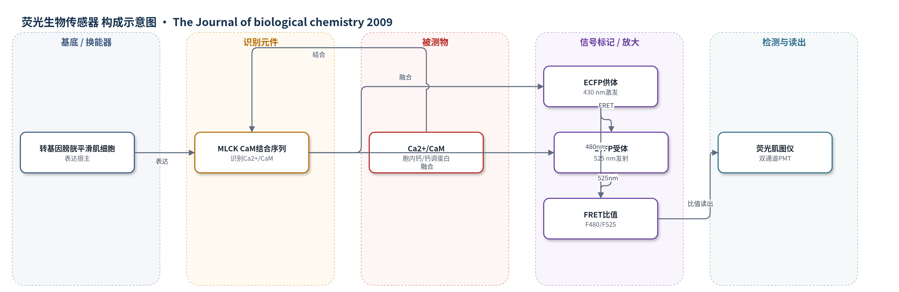
点击图片查看大图 · 依据论文自动绘制
传感器类型
荧光生物传感器
检测对象
Ca2+/钙调蛋白复合物(Ca2+/calmodulin, Ca2+/CaM)、MLCK激活状态(MLCK activation);样品基质:小鼠膀胱平滑肌组织条(urinary bladder smooth muscle strips),生理盐溶液中神经电刺激
检测原理
该传感器为基因编码FRET生物传感器:短平滑肌MLCK的CaM结合序列连接ECFP供体与EYFP受体。静息或低Ca2+时,传感器呈二聚/近距离构象,ECFP向EYFP发生显著FRET;神经刺激释放乙酰胆碱和ATP,使膀胱平滑肌[Ca2+]i升高,Ca2+结合CaM后形成Ca2+/CaM,结合MLCK CaM结合序列,导致二聚体解离和供受体距离增大,FRET强度下降。430 nm激发下,480 nm与525 nm发射比值F480/F525随Ca2+/CaM结合和MLCK激活增加而降低,从而实时反映激酶激活。该过程无酶催化沉积或核酸放大,主要依赖蛋白构象变化与FRET距离敏感性。
检测灵敏度
未报告LOD、线性范围或灵敏度斜率;时间响应上升段一相指数拟合 r^2 = 0.99
效应效果
转基因小鼠中CaM-sensor MLCK表达量低于内源激酶30%,张力潜伏期与野生型无差异,无明显表型异常。神经刺激下,阿托品抑制收缩57±7%,嘌呤拮抗剂抑制28±5%,联合抑制93±2%;神经反应为卡巴胆碱最大反应的1.45±0.18倍。[Ca2+]i、MLCK激活和RLC磷酸化半最大时间约250 ms,张力为1200 ms;RLC磷酸化由0.11±0.08升至0.51±0.06 mol P/mol RLC。舒张期[Ca2+]i、MLCK激活和张力半衰期分别为2.6±0.4、0.7±0.9和3.2±0.6 s。3 s内MYPT1、CPI-17和paxillin磷酸化基本不变,支持Ca2+/MLCK/RLC磷酸化是启动收缩的核心通路。
传感器的构成
- 表达宿主/基底:转基因小鼠膀胱平滑肌细胞,特异性表达CaM-sensor MLCK,提供生理表达环境
- 识别元件:短平滑肌MLCK的钙调蛋白结合序列(CaM-binding sequence),结合Ca2+/CaM并触发构象变化
- 换能器/荧光对:ECFP-EYFP FRET对,将CaM结合引起的距离变化转换为FRET信号
- 供体荧光标记:增强型青色荧光蛋白(ECFP),430 nm激发
- 受体荧光标记:增强型黄色荧光蛋白(EYFP),525 nm发射
- 构象连接元件:MLCK CaM结合序列连接ECFP与EYFP,Ca2+/CaM结合使二聚体解离并降低FRET
- 信号读出:荧光肌图仪(fluorescence myograph),双通道检测480/525 nm发射并计算F480/F525比值
中文摘要
本研究确定参与Ca2+/钙调蛋白(CaM)依赖性平滑肌肌球蛋白调节轻链(RLC)磷酸化的信号过程关系。作者在转基因小鼠平滑肌中表达基因编码生物传感器MLCK,实时测量神经刺激3 s的小鼠膀胱平滑肌条中[Ca2+]i、MLCK激活与张力。[Ca2+]i和激酶激活潜伏期分别为55±8 ms和65±6 ms,二者在100 ms随RLC磷酸化升高,张力潜伏期为109±3 ms。[Ca2+]i、激酶激活和RLC磷酸化于1.2 s达最大,张力于3 s达最大。MLCK激活在3 s部分下降而[Ca2+]i不变,舒张期下降更快,可能因MLCK钙调蛋白结合区磷酸化导致Ca2+脱敏。MYPT1、CPI-17和paxillin磷酸化在张力形成期未改变。结果表明神经刺激快速升高[Ca2+]i、MLCK激活和RLC磷酸化,紧密偶联的Ca2+信号复合物是启动收缩的基本机制。
英文摘要
Relationships among biochemical signaling processes involved in Ca2+/calmodulin (CaM)-dependent phosphorylation of smooth muscle myosin regulatory light chain (RLC) by myosin light chain kinase (MLCK) were determined. A genetically-encoded biosensor MLCK for measuring Ca(2+)-dependent CaM binding and activation was expressed in smooth muscles of transgenic mice. We performed real-time evaluations of the relationships among [Ca2+](i), MLCK activation, and contraction in urinary bladder smooth muscle strips neurally stimulated for 3 s. Latencies for the onset of [Ca2+](i) and kinase activation were 55 +/- 8 and 65 +/- 6 ms, respectively. Both increased with RLC phosphorylation at 100 ms, whereas force latency was 109 +/- 3 ms. [Ca2+](i), kinase activation, and RLC phosphorylation responses were maximal by 1.2 s, whereas force increased more slowly to a maximal value at 3 s. A delayed temporal response between RLC phosphorylation and force is probably due to mechanical effects associated with elastic elements in the tissue. MLCK activation partially declined at 3 s of stimulation with no change in [Ca2+](i) and also declined more rapidly than [Ca2+](i) during relaxation. The apparent desensitization of MLCK to Ca2+ activation appears to be due to phosphorylation in its calmodulin binding segment. Phosphorylation of two myosin light chain phosphatase regulatory proteins (MYPT1 and CPI-17) or a protein implicated in strengthening membrane adhesion complexes for force transmission (paxillin) did not change during force development. Thus, neural stimulation leads to rapid increases in [Ca2+](i), MLCK activation, and RLC phosphorylation in phasic smooth muscle, showing a tightly coupled Ca2+ signaling complex as an elementary mechanism initiating contraction.