组成图示
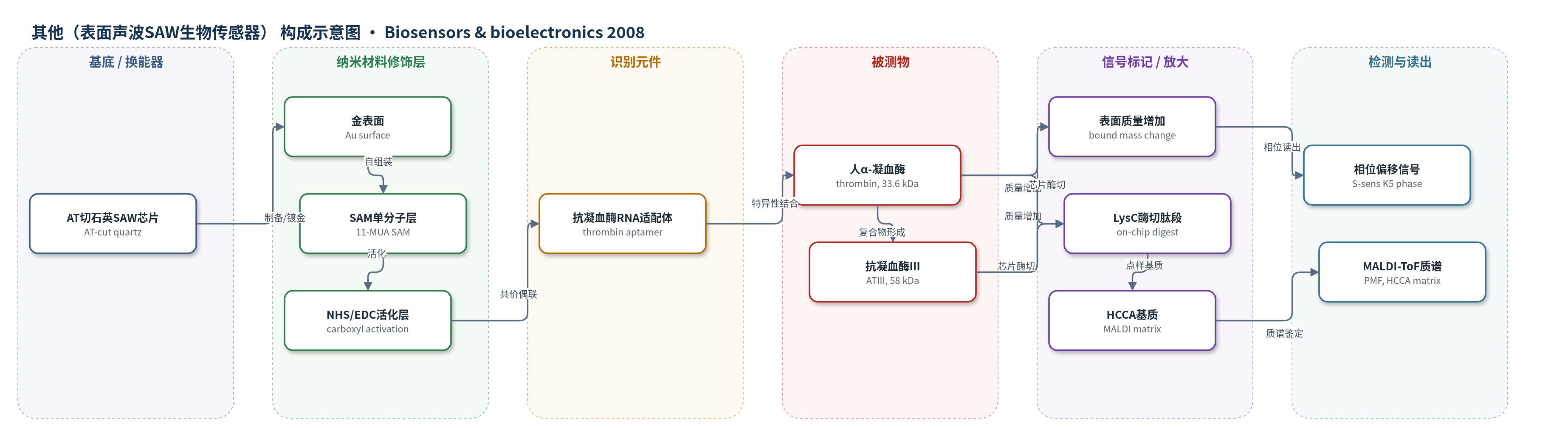
点击图片查看大图 · 依据论文自动绘制
传感器类型
其他(表面声波SAW生物传感器)
检测对象
人 α-凝血酶(human α-thrombin, thrombin)、人抗凝血酶 III(human antithrombin III, ATIII)及凝血酶–抗凝血酶 III 复合物(TAT-complex);样品基质:结合缓冲液(20 mM Tris-HCl, pH 7.4, 140 mM NaCl, 5 mM KCl, 1 mM MgCl2)
检测原理
传感器以金表面 SAM 共价固定抗凝血酶 RNA 适配体作为识别层。当结合缓冲液中的人 α-凝血酶流过芯片时,适配体特异性捕获凝血酶,使 Love 波 SAW 芯片表面质量增加;随后加入的抗凝血酶 III 与已捕获凝血酶结合形成 TAT 复合物,进一步增加表面质量。SAW 换能器将质量/黏度变化转换为相位偏移,参考元件用于扣除非特异吸附,信号随结合蛋白量近似线性变化。随后将芯片上捕获蛋白用 LysC 酶切,肽段经 HCCA 基质 MALDI-ToF MS 进行肽质量指纹鉴定;对 TAT 复合物需先经纳升级 HPLC 分离肽段,以降低复杂度和提高鉴定显著性。
检测灵敏度
LOD: <1 pg/mm2;灵敏度: 515°(cm2/µg)
效应效果
传感器结合可重复且非特异背景低:15次测量平均相位偏移6.8±0.7°,标准差约为平均值的10%;同芯片元件标准差可低至5–7%。1 µM凝血酶结合产生7.2±0.4°或6.6±0.4°,对应约425和390 pmol/cm2;300 nM ATIII结合约75和82 pmol/cm2,复合物比例为17.7%和21.1%,拟合Kd为181 nM。MALDI-ToF MS中凝血酶MOWSE分数达107,ATIII为74,并经PSD验证;TAT复合物需纳升级HPLC分离后鉴定。作者认为SAW-MS稳健、可用于蛋白相互作用、功能蛋白质组学和微型化诊断。
传感器的构成
- 基底/换能器:AT切石英晶体(AT-cut quartz crystal)光刻制备的 Love 波 SAW 芯片,含5个传感元件,将表面质量变化转换为相位信号。
- 金属表面:金(Au)屏蔽/电极表面,作为自组装单分子层成核基底。
- 自组装单分子层:11-巯基十二烷酸(11-mercaptoundecanoic acid, SAM),在金表面形成有序层并暴露羧基。
- 化学活化层:NHS 与 EDC 活化 SAM 羧基,形成可共价偶联 RNA 适配体 5′-氨基的连接位点。
- 识别元件:5′-NH2–(CH2)3–GGG AAC AAA GCU GAA GUA CUU ACC C-3′ 抗凝血酶 RNA 适配体(anti-thrombin RNA aptamer),特异性捕获人 α-凝血酶。
- 参考/对照元件:同一芯片中2个未用于适配体捕获的传感元件,作为参考扣除非特异吸附与漂移。
- 读出系统:S-sens® K5 读出系统,实时记录相位偏移;后续 MALDI-ToF MS 用于蛋白鉴定。
中文摘要
本文报道将 S-sens® K5 表面声波(SAW)生物传感器与质谱联用(SAW-MS),用于分析由人凝血级联因子 α-凝血酶(thrombin)和血浆特异性抑制剂抗凝血酶 III(antithrombin III, ATIII)组成的蛋白复合物。利用 S-sens® K5 传感器实时记录 ATIII 与凝血酶的特异性结合过程;芯片5个传感元件中2个作为参考,其余3个修饰抗凝血酶 RNA 适配体,依次结合凝血酶和 ATIII。传感器通过表面质量变化显示约 400 fmol/cm2 凝血酶中约 20% 与分子量为其 1.7 倍的 ATIII 形成复合物。随后对芯片上适配体捕获的凝血酶及凝血酶–ATIII 复合物进行蛋白酶酶切,并用 MALDI-ToF 质谱肽质量指纹法鉴定。凝血酶可直接从芯片表面整体酶切产物中显著鉴定;而 TAT 复合物中两种蛋白的显著鉴定需先经纳升级 HPLC 分离肽段再进行 MALDI-ToF MS。SAW-MS 可用于功能蛋白质组学中的蛋白相互作用分析和微型化诊断。
英文摘要
A S-sens K5 surface acoustic wave biosensor was coupled with mass spectrometry (SAW-MS) for the analysis of a protein complex consisting of human blood clotting cascade factor alpha-thrombin and human antithrombin III, a specific blood plasma inhibitor of thrombin. Specific binding of antithrombin III to thrombin was recorded as a function of time with a S-sens K5 biosensor. Two out of five elements of the sensor chip were used as references. To the remaining three elements coated with RNA anti-thrombin aptamers, thrombin and antithrombin III were bound consecutively. The biosensor measures mass changes on the chip surface showing that 20% of about 400fmol/cm2 thrombin formed a complex with the 1.7-times larger antithrombin III. Mass spectrometry (MS) was applied to identify the bound proteins. Sensor chips with aptamer-captured (1) thrombin and (2) thrombin-antithrombin III complex (TAT-complex) were digested with proteases on the sensor element and subsequently identified by peptide mass fingerprint (PMF) with matrix assisted laser desorption/ionization time-of-flight (MALDI-ToF) mass spectrometry. A significant identification of thrombin was achieved by measuring the entire digest with MALDI-ToF MS directly from the sensor chip surface. For the significant identification of both proteins in the TAT-complex, the proteolytic peptides had to be separated by nano-capillary-HPLC prior to MALDI-ToF MS. SAW-MS is applicable to protein interaction analysis as in functional proteomics and to miniaturized diagnostics.