组成图示
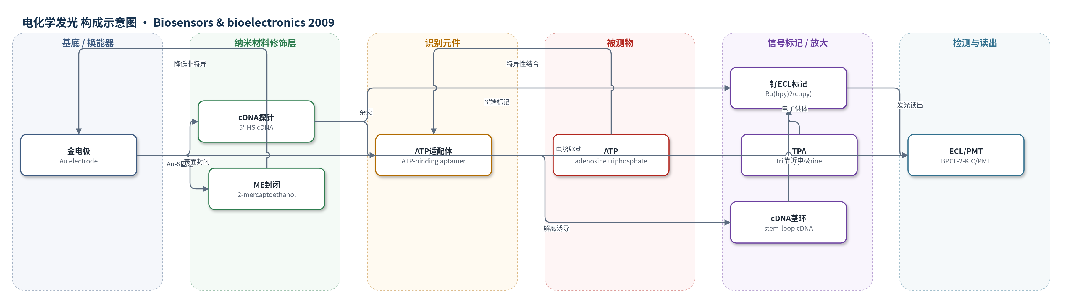
点击图片查看大图 · 依据论文自动绘制
传感器类型
电化学发光(ECL)生物传感器
检测对象
ATP(adenosine triphosphate,三磷酸腺苷);样品基质为PBS缓冲液/水溶液(10 mM PBS,0.1 M NaCl,pH 7.4),非实际生物样品
检测原理
传感器以金电极为工作电极,cDNA通过5'-HS与Au-S键固定,3'端标记Ru(bpy)2(cbpy)。初始cDNA与ATP适配体杂交形成刚性ds-DNA,钌发光体远离电极,ECL较弱。加入ATP后,适配体优先与ATP结合并从电极表面解离,cDNA仍固定于电极;随后在1 M NaCl和5 mM MgCl2高离子强度溶液中,cDNA两端互补序列形成茎环结构,使Ru(bpy)2(cbpy)靠近电极表面。在0.10 M PBS(pH 7.4)含0.10 M TPA体系中,循环伏安扫描(0–0.75 V)激发Ru配合物与TPA发生共反应,产生ECL。ATP浓度越高,解离的适配体越多,茎环cDNA比例越高,ECL越强,信号与ATP浓度对数线性相关。
检测灵敏度
LOD: 0.02 nM(S/N=3);线性范围: 0.05 nM–10 nM(ECL强度与ATP浓度对数线性);IECL = 470.6 + 151 lgC(C单位nM);相关系数: 0.994
效应效果
该传感器对ATP具有良好选择性:10 nM CTP和GTP的ECL响应与空白一致,ATP/CTP/GTP混合液中的信号与单独ATP相近,表明交叉干扰可忽略。方法重复性通过三个重复标准样品误差棒评价,但未报告RSD数值。传感器可在37℃下用1 μM ATP结合适配体溶液孵育1 h再生,无需高温;三次再生循环后平均ECL强度恢复至初始值的90%,显示可重复使用潜力。其LOD为0.02 nM,低于文中比较的先前ATP传感器报道。作者认为该cDNA探针ECL-AB体系成本低、再生温和、灵敏度高、选择性好,可作为适配体小分子检测的通用策略。
传感器的构成
- 基底电极:金电极(Au electrode),抛光与电化学预处理,作为ECL工作电极
- 探针DNA:ATP适配体互补DNA(cDNA),5'-HS端自组装固定,3'-NH2端标记Ru,两端互补碱基可形成茎环
- 识别元件:ATP结合适配体(ATP-binding aptamer),与cDNA杂交形成刚性双链,结合ATP后解离
- 信号标记物:Ru(bpy)2(cbpy)(由Ru(bpy)2(cbpy)NHS标记),共价连接cDNA 3'端,作为ECL发光体
- 封闭剂:2-巯基乙醇(ME),置换非特异性结合寡核苷酸并钝化金表面
- 电子供体:三丙胺(TPA),ECL测量中与Ru配合物共反应产生发光
- 信号放大介质:1 M NaCl/5 mM MgCl2(PBS pH 7.4),诱导cDNA形成茎环并使Ru靠近电极
中文摘要
本文报道了一种基于适配体的电化学发光(ECL)生物传感器,用于高灵敏、高选择地检测三磷酸腺苷(ATP)。传感器构建时,先将ATP结合适配体的互补DNA(cDNA)与适配体杂交,形成刚性线状双链DNA(ds-DNA);随后在cDNA的3′端共价标记钌配合物Ru(bpy)2(cbpy),并通过cDNA 5′端巯基将ds-DNA自组装固定于金电极表面,再用2-巯基乙醇封闭非特异性结合位点。检测时,ATP与适配体特异性结合,使适配体从ds-DNA上解离,cDNA在高离子强度MgCl2溶液中形成茎环结构,使钌发光体靠近电极表面,从而增强ECL信号。ECL强度与ATP浓度对数在0.05 nM至10 nM范围内线性相关,检出限为0.02 nM。该体系以cDNA作为探针,适配体无需标记,且可通过适配体溶液温和再生,为适配体小分子检测提供了可重复、简便的模型。
英文摘要
An aptamer-based electrochemiluminescent (ECL-AB) biosensor for ATP detection with high sensitivity and specificity was developed. The biosensor was assembled based on several steps. First, a complementary DNA (cDNA) of the ATP-binding aptamer, which has six complementary bases at both its ends, was hybridized with the aptamer molecule to form a rigid, linear double-stranded DNA (ds-DNA). The ds-DNA was then labeled with a ruthenium complex at the 3' terminus of cDNA, followed by the immobilization of this ds-DNA onto Au electrode surface through the 5'-HS on the cDNA. In the presence of ATP, due to the ATP binding to the aptamer, the aptamer molecules dissociated from the ds-DNA complex, which resulted in the formation of stem-loop structure of the single-stranded cDNA and led to the increase of the ECL signal. The increased ECL intensity was found linearly to the logarithm of the concentration of ATP ranging from 0.05 nM to 10 nM with a detection limit of 0.02 nM. Different from other ECL-AB biosensors with aptamers as the probes, this sensing system proposed here is based on the utilization of the cDNA of aptamers as the probes for ECL sensing. Therefore, such sensing system could provide a promising label-free and more readily regenerated model for aptamer-based small-molecules detection.