组成图示
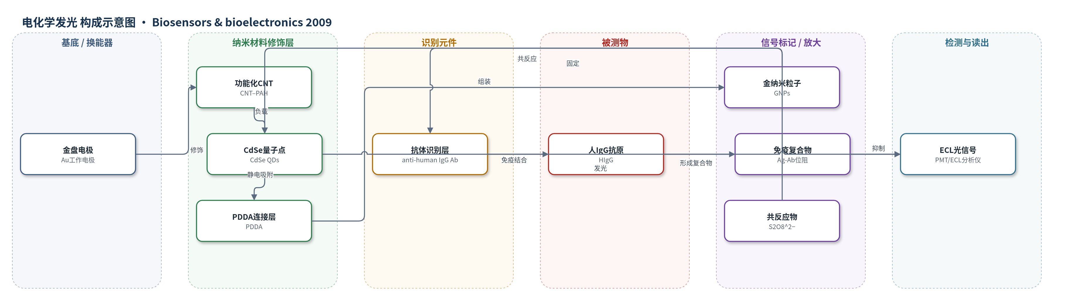
点击图片查看大图 · 依据论文自动绘制
传感器类型
电化学发光(ECL)生物传感器
检测对象
人免疫球蛋白G(human IgG,HIgG),样品基质为人血清(serum)
检测原理
在0.1 M PBS(pH 7.4)含0.1 M K2S2O8和0.1 M KCl体系中,电极上的CdSe QDs被还原为CdSe•−,S2O8^2−被还原生成强氧化剂SO4•−;二者发生电子转移产生激发态CdSe*并发出ECL光。CNTs提高复合膜导电性和孔隙率,PDDA的氨基催化ECL反应,GNPs作为导电桥并增加抗体负载,实现信号放大。固定于电极的抗人IgG抗体与目标HIgG结合后形成免疫复合物,增加界面空间位阻,阻碍共反应物向QDs扩散和电子转移,使ECL强度随HIgG浓度升高而降低,从而实现定量检测。
检测灵敏度
LOD: 0.6 pg mL−1;线性范围: 0.002–500 ng L−1
效应效果
该传感器对干扰物选择性良好:2 ng mL−1人IgG与200 ng mL−1山羊IgG、200 ng mL−1低密度脂蛋白(LDL)混合后,ECL响应与纯人IgG相比无显著差异(R.S.D=8.4%)。传感器在pH 7.4 PBS中4 ℃保存30 d后性能无明显下降。四个同批制备传感器检测20 ng mL−1 IgG的批间RSD为8.3%。经0.2 M甘氨酸盐酸盐(pH 2.8)处理9 min可再生,20 ng mL−1样品内检CV为9.8%。人血清样品与ELISA结果一致:0.032/0.03、0.40/0.45、24.4/23.0 ng mL−1,相对偏差分别为4.56%、−8.3%、4.2%,表明其可用于临床血清IgG检测。
传感器的构成
- 基底/换能器电极:金盘电极(Au disk electrode),作为工作电极承载修饰层并传递电子
- 纳米材料修饰层:PAH功能化多壁碳纳米管(CNT–PAH)负载CdSe量子点(CdSe QDs),形成CdSe QDs–CNTs复合膜,增强ECL并提高导电性
- 聚合物连接/增强层:聚二烯丙基二甲基氯化铵(PDDA)静电吸附于复合膜,作为连接链并催化增强ECL
- 信号放大/抗体固定层:金纳米粒子(GNPs)组装于PDDA层,作为导电桥并固定抗体、放大ECL
- 识别元件:山羊抗人IgG抗体(goat anti-human IgG Ab),特异性结合人IgG抗原
- 封闭剂:牛血清白蛋白(BSA),封闭非特异性结合位点
- 电解质/共反应物:0.1 M PBS(pH 7.4)、0.1 M K2S2O8、0.1 M KCl,提供反应介质与ECL共反应物
中文摘要
本文报道了一种基于CdSe量子点(QDs)与碳纳米管(CNTs)及聚二烯丙基二甲基氯化铵(PDDA)复合膜的电化学发光(ECL)免疫传感器,用于灵敏检测人免疫球蛋白G(human IgG,HIgG)。CdSe QDs–CNTs复合材料具有较高ECL强度、良好生物相容性和稳定性。将PDDA作为连接链结合到电极上的CdSe QDs–CNTs复合膜后,ECL信号显著增强;随后在CdSe QDs–CNTs/PDDA修饰电极上组装金纳米粒子(GNPs),可再次放大ECL信号。抗体(Ab)通过GNPs固定在电极表面,完成免疫传感器构建。该工作首次探索了PDDA增强QDs ECL的独特功能。检测原理基于免疫反应后空间位阻增加,使ECL强度降低。人IgG在0.002–500 ng L−1范围内呈线性,检出限为0.6 pg mL−1。传感器制备与检测重现性良好,临床血清检测结果与现有方法一致,具有临床应用前景。
英文摘要
Electrochemiluminescence (ECL) of CdSe quantum dots (QDs) was greatly enhanced by the combination of carbon nanotubes (CNTs) and poly (diallyldimethylammonium chloride) (PDDA) in the CdSe QDs film, and could successfully be used to develop a sensitive ECL immunosensor for the detection of human IgG (Ag). The novel CdSe QDs-CNTs composites exhibited high ECL intensity, good biocompatibility, and high stability, which held great promise for the fabrication of the ECL biosensors with improved sensitivity. After PDDA as a binding linker was conjugated to the CdSe QDs-CNTs composite film on the electrode, the ECL signal was significantly enhanced. Subsequently, gold nanoparticles (GNPs) assembled onto the CdSe QDs-CNTs/PDDA modified electrode could amplify the ECL signal once again. After antibody (Ab) was immobilized onto the electrode through GNPs, the ECL immunosensor was successfully fabricated. It is for the first time that the unique function of PDDA for enhancing QDs ECL was explored and used to develop an ECL biosensor. The principle of ECL detection for target Ag is based on the increment of steric hindrance after immunoreaction, which resulted in the decrease of ECL intensity. The Ag concentration was determined in the linear range of 0.002-500 ng L(-1) with a detection limit of 0.6 pg mL(-1). The sensor showed good fabrication and detection reproducibility, and the assay results were in acceptable agreement with the clinical sera tests, showing a promising clinical application. This work opened the new avenues for applying QDs ECL in highly sensitive bioassays.