组成图示
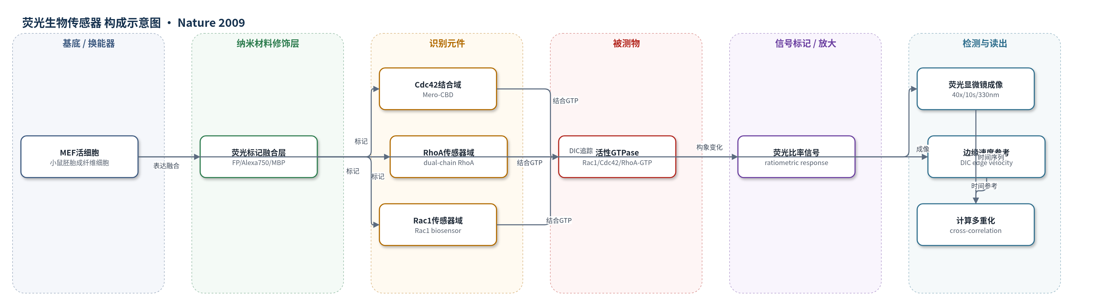
点击图片查看大图 · 依据论文自动绘制
传感器类型
荧光生物传感器
检测对象
活性 Rac1(Rac1-GTP)、活性 Cdc42(Cdc42-GTP)、活性 RhoA(RhoA-GTP);样品基质:小鼠胚胎成纤维细胞(MEF)活细胞,细胞前沿/细胞质
检测原理
GTPase 荧光生物传感器包含可识别活性 GTPase-GTP 的结合域和荧光报告基团。当 Rac1、Cdc42 或 RhoA 与 GTP 结合并被激活时,传感器结合域发生构象变化,改变荧光蛋白或 Alexa750 染料之间的荧光比率,从而将分子激活状态转换为可成像的荧光信号。活细胞以 10 s 间隔进行比率荧光成像,并经体积、串扰和传感器浓度校正。由于不同 GTPase 传感器不能同时表达,方法以细胞边缘突出/回缩速度作为共同时间参考,将不同实验中的激活时间序列对齐,通过 Pearson 互相关和 bootstrap 平均提取各 GTPase 激活相对于边缘运动的时滞与空间位置。局部采样窗口和多细胞平均用于降低噪声并提高统计可靠性。
检测灵敏度
原文未报告 LOD、线性范围、灵敏度斜率或 R^2。
效应效果
该方法在 MEF 中表现出可重复的时空分辨:RhoA 激活与边缘前进同步,且与突出相关活性限制在距前沿约 2 μm 内;Rac1 和 Cdc42 峰值激活滞后约 40 s,初始激活分别位于距前沿 1.8±0.7 μm 和 1.3±0.7 μm。跨细胞 bootstrap 分析给出时滞:Rac1 -41 s(95% CI -61 至 -13),Cdc42 -46 s(-51 至 -39),RhoA -6 s(-8 至 -4)。直接双通道 Cdc42/RhoA 成像得到 Cdc42 滞后 RhoA -34 s(-30 至 -38,n=7),与计算多重化预测一致。控制实验表明不同传感器设计和表达量不改变相对激活动力学。作者主张该方法可低扰动、可扩展地重建多信号分子协调。
传感器的构成
- 细胞基底:小鼠胚胎成纤维细胞(MEF)活细胞,提供 GTPase 激活与细胞突出微环境
- 识别元件:Cdc42 生物传感器结合域(Mero-CBD),识别活性 Cdc42-GTP
- 识别元件:RhoA 双链生物传感器(dual-chain RhoA biosensor),识别活性 RhoA-GTP
- 识别元件:Rac1 荧光蛋白型生物传感器(Rac1 biosensor),识别活性 Rac1-GTP
- 信号标记物:Alexa750 染料与荧光蛋白(FP),用于比率荧光报告
- 辅助融合:麦芽糖结合蛋白(MBP),融合于 Cdc42 传感器以提高溶解度
- 换能读出:荧光显微镜比率成像(40×/1.3 NA,10 s 间隔,330 nm 像素)与 DIC 边缘速度追踪
中文摘要
Rac1、RhoA 和 Cdc42 等 Rho 家族 GTPase 协同调控细胞骨架动态,但它们在单个细胞突出事件中秒级、微米级的时空协调仍不清楚。本研究在小鼠胚胎成纤维细胞(MEF)中,利用两种 GTPase 荧光生物传感器的同步成像,以及一种以突出/回缩事件为共同时间参考的“计算多重化”方法,分别测定 Rac1、Cdc42 和 RhoA 的激活动力学。结果显示,RhoA 在细胞边缘与边缘前进同步激活,而 Cdc42 和 Rac1 在距边缘约 2 μm 处滞后约 40 s 激活。这表明 Rac1 与 RhoA 通过空间分离和精确时序发挥拮抗作用;RhoA 参与突出的初始事件,Rac1 和 Cdc42 则激活与新生突出强化和稳定相关的通路。计算多重化方法可在不同实验间对齐多个蛋白活性,为低扰动重建细胞信号通路提供工具。
英文摘要
The GTPases Rac1, RhoA and Cdc42 act together to control cytoskeleton dynamics. Recent biosensor studies have shown that all three GTPases are activated at the front of migrating cells, and biochemical evidence suggests that they may regulate one another: Cdc42 can activate Rac1 (ref. 8), and Rac1 and RhoA are mutually inhibitory. However, their spatiotemporal coordination, at the seconds and single-micrometre dimensions typical of individual protrusion events, remains unknown. Here we examine GTPase coordination in mouse embryonic fibroblasts both through simultaneous visualization of two GTPase biosensors and using a 'computational multiplexing' approach capable of defining the relationships between multiple protein activities visualized in separate experiments. We found that RhoA is activated at the cell edge synchronous with edge advancement, whereas Cdc42 and Rac1 are activated 2 micro-m behind the edge with a delay of 40 s. This indicates that Rac1 and RhoA operate antagonistically through spatial separation and precise timing, and that RhoA has a role in the initial events of protrusion, whereas Rac1 and Cdc42 activate pathways implicated in reinforcement and stabilization of newly expanded protrusions.