组成图示
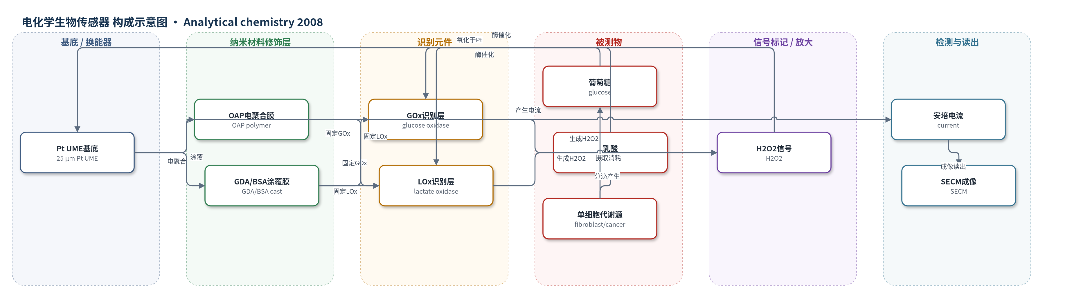
点击图片查看大图 · 依据论文自动绘制
传感器类型
电化学生物传感器
检测对象
葡萄糖(glucose)、乳酸(lactate);样品基质为单个活细胞(成纤维细胞、MCF10CA1a 癌细胞、PC12 细胞)上方细胞外培养液(HBSS 或 RPMI)
检测原理
该传感器为酶促安培型。GOx 催化 D-葡萄糖与 O2 生成 D-葡萄糖内酯和 H2O2;LOx 催化 L-乳酸与 O2 生成丙酮酸和 H2O2。酶被 OAP 电聚合膜或 GDA/BSA 涂覆膜固定于 Pt UME 表面,形成选择性识别层。底物在酶层内反应生成的 H2O2 扩散至 Pt 表面,在 +0.6 V(vs Ag/AgCl)发生氧化,产生与底物浓度相关的安培电流。细胞摄取葡萄糖使局部葡萄糖降低,电流下降;细胞分泌乳酸使局部乳酸升高,电流上升。SECM 恒高扫描时,电流空间分布即可映射单细胞代谢。酶膜同时阻挡多巴胺、外源 H2O2 等干扰,低浓度乳酸区符合 Michaelis-Menten 动力学。
检测灵敏度
线性范围: GOx 电聚合 0–18 mM;GOx 涂覆 0–14 mM;LOx 电聚合 0.1–0.3 mM 与 0.4–0.9 mM(>0.9 mM 灵敏度下降);LOx 涂覆 0.1–0.3 mM 与 0.4–1.0 mM
效应效果
传感器依靠酶膜选择性,聚合物膜可阻挡干扰;MAMP 活细胞测试未发现交叉干扰。稳定性:电聚合 GOx 4 °C 保存 12 天几乎不变,可保持约 1 个月;涂覆 GOx 室温 2–3 天后下降。LOx 电聚合约 24 h,涂覆约 2 天。重现性:电聚合优于涂覆,相隔数月制备的电聚合 GOx 接近曲线相似。制造成功率:GOx 电聚合约 70%、涂覆约 90%;LOx 电聚合约 20%、涂覆约 60%。单细胞实验中,无细胞背景无乳酸电流增加;8 µm 氧呼吸显示细胞接触处活性更高。用于单细胞代谢成像。
传感器的构成
- 换能器电极:25 µm Pt UME,铂超微电极,氧化 H2O2 并输出安培电流
- 电聚合修饰层:OAP(2-氨基苯酚)聚合物膜,电聚合固定 GOx/LOx
- 涂覆修饰层:GDA(戊二醛)与 BSA(牛血清白蛋白)混合膜,手工涂覆固定 GOx/LOx 并封闭/稳定
- 识别/催化元件:GOx 或 LOx,分别催化葡萄糖或乳酸氧化生成 H2O2
- 信号产物:H2O2,酶催化生成并在 Pt UME 表面氧化
中文摘要
本文报道了用于扫描电化学显微镜(SECM)成像单个活细胞的葡萄糖和乳酸超微电极(UME)生物传感器。传感器以铂 UME 为换能器,分别通过电聚合或手工涂覆将葡萄糖氧化酶(GOx)和乳酸氧化酶(LOx)固定于电极表面,并测定其对葡萄糖和乳酸的安培响应。评价表明,两种制备方式各有优势:手工涂覆具有更高灵敏度和更短制造时间,电聚合则具有更好重现性。作者获得了各类生物传感器 UME 的安培接近曲线(AC),可用于将 UME 定位在基底上方已知距离。利用葡萄糖生物传感器 UME,在单个成纤维细胞上方记录了葡萄糖摄取轮廓;利用乳酸生物传感器 UME,在单个癌细胞上方记录了乳酸产生轮廓。此外,单个癌细胞的氧呼吸轮廓并不简单复制细胞形貌,而更为复杂,在细胞与培养皿接触处观察到更高呼吸活性。这些 UME 生物传感器有望成为绘制单癌细胞葡萄糖、乳酸和氧等代谢分析物分布的有力工具。
英文摘要
We have developed glucose and lactate ultramicroelectrode (UME) biosensors based on glucose oxidase and lactate oxidase (with enzymes immobilized onto Pt UMEs by either electropolymerization or casting) for scanning electrochemical microscopy (SECM) and have determined their sensitivity to glucose and lactate, respectively. The results of our evaluations reveal different advantages for sensors constructed by each method: improved sensitivity and shorter manufacturing time for hand-casting, and increased reproducibility for electropolymerization. We have acquired amperometric approach curves (ACs) for each type of manufactured biosensor UME, and these ACs can be used as a means of positioning the UME above a substrate at a known distance. We have used the glucose biosensor UMEs to record profiles of glucose uptake above individual fibroblasts. Likewise, we have employed the lactate biosensor UMEs for recording the lactate production above single cancer cells with the SECM. We also show that oxygen respiration profiles for single cancer cells do not mimic cell topography, but are rather more convoluted, with a higher respiration activity observed at the points where the cell touches the Petri dish. These UME biosensors, along with the application of others already described in the literature, could prove to be powerful tools for mapping metabolic analytes, such as glucose, lactate, and oxygen, in single cancer cells.