组成图示
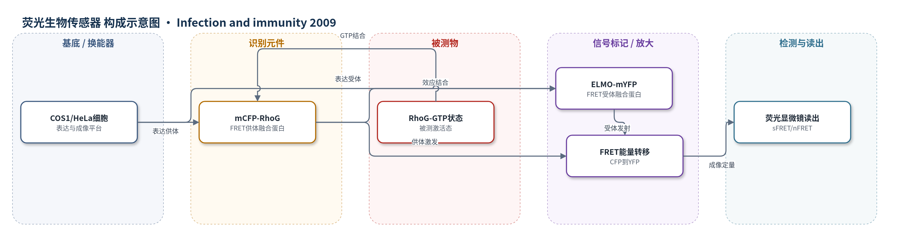
点击图片查看大图 · 依据论文自动绘制
传感器类型
荧光生物传感器
检测对象
RhoG 激活状态(RhoG activation state,GTP 结合态 RhoG);样品基质:COS1/HeLa 细胞胞质与细胞膜
检测原理
该 FRET 生物传感器由 mCFP-RhoG 和 ELMO(AA1-362)-mYFP 两个融合蛋白组成。RhoG 结合 GTP 后构象改变,与 ELMO 氨基端 1–362 氨基酸特异性结合,使 mCFP 与 mYFP 距离缩短至 FRET 有效范围;CFP 受激发后能量非辐射转移至 YFP,产生 YFP 发射。RhoG-GTP 比例越高,供体-受体偶联越多,FRET 信号越强;GDP 态、T17N 或效应结合突变 F37A/Y40C 降低结合,FRET 信号下降。通过 CFP、YFP 和 FRET 通道成像,扣除串扰与交叉激发后计算 sFRET 和 nFRET,即可在单细胞水平空间分辨 RhoG 激活。YopE 使 RhoG 水解 GTP 而降低局部 FRET,YopT 切割 C 端异戊二烯化位点导致 RhoG 错误定位并改变局部信号。
检测灵敏度
—
效应效果
该 FRET 传感器对 RhoG 核苷酸状态敏感:G12V 构象的 FRET 高于野生型,T17N 显著降低,效应结合突变 F37A/Y40C 也降低;sFRET 中野生型对 G12V P<0.0001、对 T17N P=0.0007,nFRET 中 P<0.05 和 P=0.0024。细菌附着位点 RhoG 激活显著升高,sFRET P<0.0001、nFRET P=0.0185;Rac1 对照 sFRET P=0.0004、nFRET P=0.0014。YopE 使附着位点 RhoG FRET 降低(P=0.0053),但无细菌背景基本不变(P=0.883)。YopT 使 RhoG 从新生吞噬体减少(P<0.0001),Rac1 亦减少(P=0.0021),非底物 Arf6 无变化(P=0.464)。作者主张该传感器可空间分辨监测 RhoG 激活及耶尔森菌效应蛋白调控。
传感器的构成
- 表达基底:COS1/HeLa 细胞,作为 FRET 生物传感器的表达与成像平台,提供胞质和细胞膜微环境
- 表达载体:pmCFP-C1 与 pmYFP-N1 质粒,分别携带 mCFP-RhoG 和 ELMO(AA1-362)-mYFP 构建体,用于转染表达
- 供体识别元件:mCFP-RhoG,mCFP 融合 RhoG,作为 FRET 供体并承载 GTPase 核苷酸状态
- 受体信号元件:ELMO(AA1-362)-mYFP,ELMO2 氨基端 1–362 氨基酸融合 mYFP,作为 FRET 受体,仅在 RhoG-GTP 结合时靠近供体
- 识别机制:RhoG 与 ELMO(AA1-362) 的 GTP 依赖性相互作用,用于识别 RhoG 激活态
- 读出装置:荧光显微镜/FRET 成像系统,采集 CFP、YFP 和 FRET 通道并计算 sFRET/nFRET
中文摘要
假结核耶尔森菌表面蛋白 invasin 以高亲和力结合多种 β1 整合素,导致 Rac1 活性失调。细菌与宿主细胞结合后,多种经 III 型分泌系统转入胞质的耶尔森菌外膜蛋白(Yops)进一步改变 Rho GTPase 活性。本研究报道该菌三种毒力因子调控 Rho GTPase RhoG。细菌结合使 RhoG 被强烈募集至附着位点,且依赖 invasin 与 β1 整合素的高亲和力结合;抑制 RhoG 活性显著降低 invasin 介导的细菌内化效率。为检测 RhoG 激活状态,作者构建了基于荧光共振能量转移(FRET)的激活生物传感器,并显示 RhoG 在细菌附着位点发生空间特异性激活。该传感器还证明 YopE 作为强效 Rho GAP 可高效使 RhoG 失活;两种独立实验进一步证明异戊二烯半胱氨酸内切蛋白酶 YopT 使 RhoG 错误定位。功能摄取实验表明,RhoG 激活可弥补 Rac1 活性缺陷;增大颗粒尺寸后结果更符合 RhoG 作为 Rac1 上游激活因子的线性通路,提示吞噬表面面积增加会限制信号通路。综上,多种毒力因子共同失调 RhoG,可能通过干扰中性粒细胞功能抑制宿主免疫。
英文摘要
The Yersinia pseudotuberculosis surface protein invasin binds to multiple beta1 integrins with high affinity, leading to misregulation of Rac1 activity. Upon host cell binding, alteration of Rho GTPase activity results from the action of several Yersinia outer proteins (Yops) that are translocated into the cytoplasm. We report here that three virulence determinants encoded by Y. pseudotuberculosis manipulate the Rho GTPase RhoG. Y. pseudotuberculosis binding to cells caused robust recruitment of RhoG to the site of attachment, which required high-affinity invasin-beta1 integrin association. Furthermore, inactivation of RhoG significantly reduced the efficiency of invasin-mediated bacterial internalization. To investigate the activation state of RhoG, a fluorescence resonance energy transfer-based activation biosensor was developed and used to show distinct spatial activation of RhoG at the site of bacterial attachment. The biosensor was also used to show efficient RhoG inactivation by Y. pseudotuberculosis YopE, a potent Rho GTPase activating protein. Additionally, RhoG mislocalization by the prenylcysteine endoprotease YopT was demonstrated by two independent assays. Functional bacterial uptake experiments demonstrated that RhoG activation can bypass a deficit in Rac1 activity. Interestingly, increasing the size of the particle gave results more consistent with a linear pathway, in which RhoG acts as an upstream activator of Rac1, indicating that increased surface area introduces constraints on the signaling pathways required for efficient internalization. Taken together, these data demonstrate the misregulation of RhoG by multiple Y. pseudotuberculosis virulence determinants. Since RhoG is imperative for proper neutrophil function, this misregulation may represent a unique mechanism by which Yersinia species dampen the immune response.